Institute of Physiological Chemistry and Pathobiochemistry, University of Muenster, Muenster, Germany.
Cells-in-Motion Interfaculty Centre, University of Muenster, Muenster, Germany.
Diabetologia. 2021 Jul;64(7):1626-1641. doi: 10.1007/s00125-021-05453-z. Epub 2021 Apr 29.
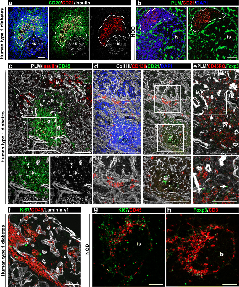
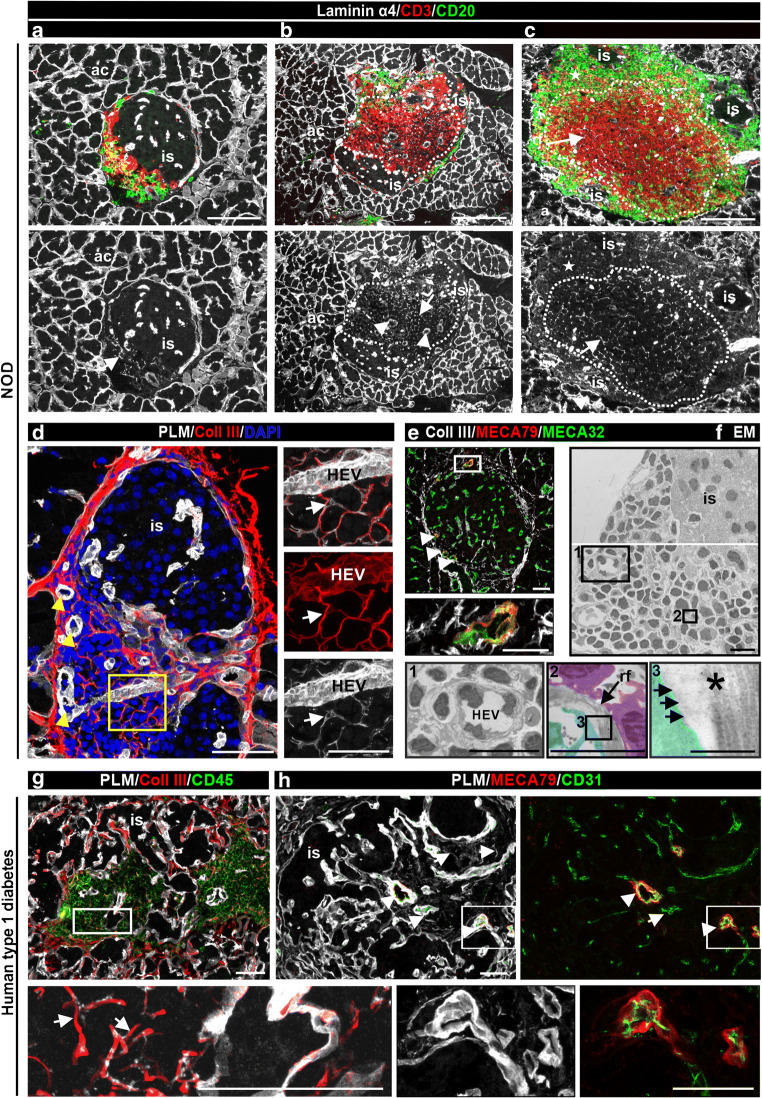
AIMS/HYPOTHESIS: We and others previously reported the presence of tertiary lymphoid organs (TLOs) in the pancreas of NOD mice, where they play a role in the development of type 1 diabetes. Our aims here are to investigate whether TLOs are present in the pancreas of individuals with type 1 diabetes and to characterise their distinctive features, in comparison with TLOs present in NOD mouse pancreases, in order to interpret their functional significance.
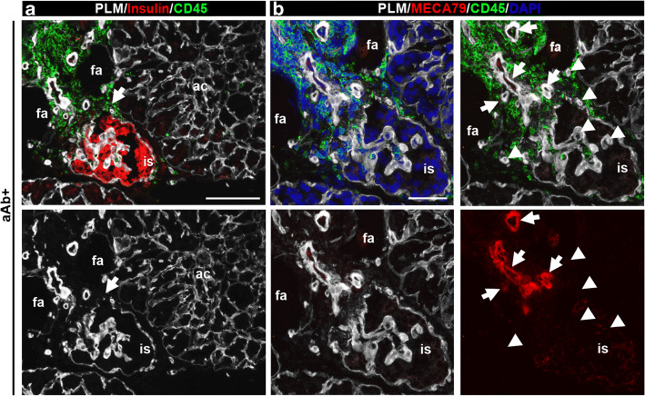
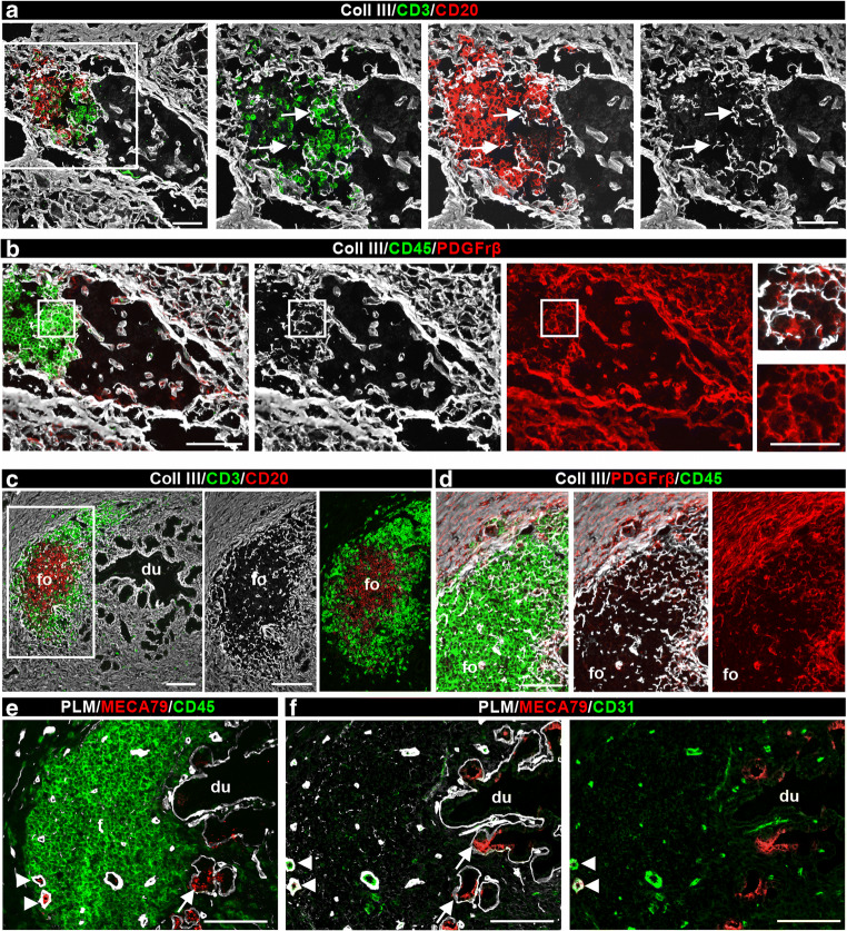
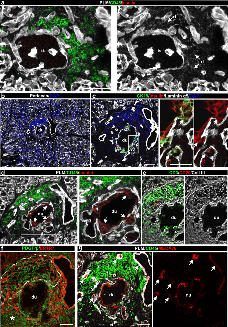
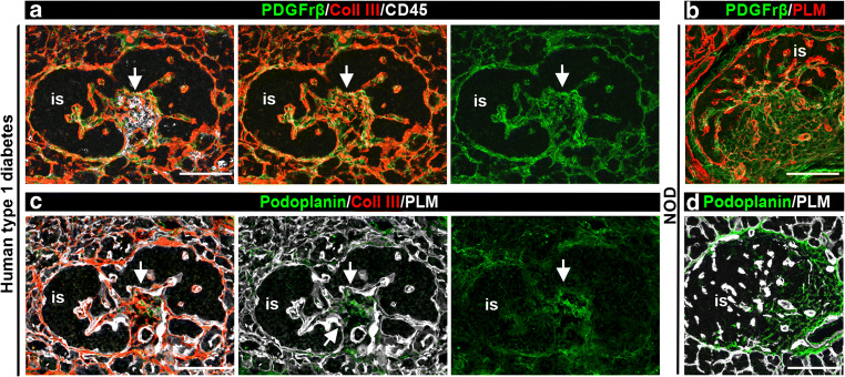
Using immunofluorescence confocal microscopy, we examined the extracellular matrix (ECM) and cellular constituents of pancreatic TLOs from individuals with ongoing islet autoimmunity in three distinct clinical settings of type 1 diabetes: at risk of diabetes; at/after diagnosis; and in the transplanted pancreas with recurrent diabetes. Comparisons were made with TLOs from 14-week-old NOD mice, which contain islets exhibiting mild to heavy leucocyte infiltration. We determined the frequency of the TLOs in human type 1diabetes with insulitis and investigated the presence of TLOs in relation to age of onset, disease duration and disease severity.
TLOs were identified in preclinical and clinical settings of human type 1 diabetes. The main characteristics of these TLOs, including the cellular and ECM composition of reticular fibres (RFs), the presence of high endothelial venules and immune cell subtypes detected, were similar to those observed for TLOs from NOD mouse pancreases. Among 21 donors with clinical type 1 diabetes who exhibited insulitis, 12 had TLOs and had developed disease at younger age compared with those lacking TLOs. Compartmentalised TLOs with distinct T cell and B cell zones were detected in donors with short disease duration. Overall, TLOs were mainly associated with insulin-containing islets and their frequency decreased with increasing severity of beta cell loss. Parallel studies in NOD mice further revealed some differences in so far as regulatory T cells were essentially absent from human pancreatic TLOs and CCL21 was not associated with RFs.
CONCLUSIONS/INTERPRETATION: We demonstrate a novel feature of pancreas pathology in type 1 diabetes. TLOs represent a potential site of autoreactive effector T cell generation in islet autoimmunity and our data from mouse and human tissues suggest that they disappear once the destructive process has run its course. Thus, TLOs may be important for type 1 diabetes progression.
目的/假设:我们和其他人之前曾报道过在 NOD 小鼠的胰腺中存在三级淋巴器官(TLOs),它们在 1 型糖尿病的发展中发挥作用。我们的目的是研究 TLOs 是否存在于 1 型糖尿病患者的胰腺中,并描述其与 NOD 小鼠胰腺 TLOs 的特征区别,以便解释其功能意义。
我们使用免疫荧光共聚焦显微镜,在 1 型糖尿病的三种不同临床环境中,即糖尿病风险、诊断时和移植后复发糖尿病的患者中,检查正在发生胰岛自身免疫的个体的胰腺 TLO 的细胞外基质(ECM)和细胞成分。将这些结果与 14 周龄 NOD 小鼠的 TLO 进行比较,后者的胰岛存在从轻到重的白细胞浸润。我们确定了人类 1 型糖尿病伴胰岛炎中 TLO 的频率,并研究了 TLO 的存在与发病年龄、疾病持续时间和疾病严重程度的关系。
在人类 1 型糖尿病的临床前和临床环境中都发现了 TLOs。这些 TLO 的主要特征,包括网状纤维(RFs)的细胞和 ECM 组成、高内皮静脉和检测到的免疫细胞亚型的存在,与 NOD 小鼠胰腺 TLOs 的特征相似。在 21 名具有胰岛炎的临床 1 型糖尿病供体中,有 12 名有 TLOs,并且发病年龄较无 TLOs 的患者更早。在疾病持续时间较短的供体中,检测到具有独特 T 细胞和 B 细胞区室的分隔 TLOs。总体而言,TLOs 主要与含有胰岛素的胰岛有关,其频率随着β细胞丢失程度的增加而降低。在 NOD 小鼠中的平行研究进一步揭示了一些差异,即人类胰腺 TLOs 中基本不存在调节性 T 细胞,而 CCL21 与 RFs 无关。
结论/解释:我们展示了 1 型糖尿病中胰腺病理学的一个新特征。TLOs 代表胰岛自身免疫中自身反应性效应 T 细胞产生的潜在部位,我们来自小鼠和人类组织的研究数据表明,一旦破坏性过程进行,它们就会消失。因此,TLOs 可能对 1 型糖尿病的进展很重要。