• 文献检索
  • 文档翻译
  • 深度研究
  • 学术资讯
  • Suppr Zotero 插件Zotero 插件
  • 邀请有礼
  • 套餐&价格
  • 历史记录
应用&插件
Suppr Zotero 插件Zotero 插件浏览器插件Mac 客户端Windows 客户端微信小程序
定价
高级版会员购买积分包购买API积分包
服务
文献检索文档翻译深度研究API 文档MCP 服务
关于我们
关于 Suppr公司介绍联系我们用户协议隐私条款
关注我们

Suppr 超能文献

核心技术专利:CN118964589B侵权必究
粤ICP备2023148730 号-1Suppr @ 2026

文献检索

告别复杂PubMed语法,用中文像聊天一样搜索,搜遍4000万医学文献。AI智能推荐,让科研检索更轻松。

立即免费搜索

文件翻译

保留排版,准确专业,支持PDF/Word/PPT等文件格式,支持 12+语言互译。

免费翻译文档

深度研究

AI帮你快速写综述,25分钟生成高质量综述,智能提取关键信息,辅助科研写作。

立即免费体验

对非重症 COVID-19 阳性患者的眼底评估。

Fundus evaluation in COVID-19 positives with non-severe disease.

作者信息

Bypareddy Ravi, Rathod B L Sujatha, Shilpa Y D, Hithashree H R, Nagaraj Kalpana Badami, Hemalatha B C, Basumatary Jessica, Bekal Deeksha, Niranjan R, Anusha P G

机构信息

Department of Vitreo-Retina, Regional Institute of Ophthalmology, Minto Ophthalmic Hospital, BMCRI, Bengaluru, Karnataka, India.

出版信息

Indian J Ophthalmol. 2021 May;69(5):1271-1274. doi: 10.4103/ijo.IJO_3227_20.

DOI:10.4103/ijo.IJO_3227_20
PMID:33913875
原文链接:https://pmc.ncbi.nlm.nih.gov/articles/PMC8186568/
Abstract

PURPOSE

The aim of this work was to study and document retinal changes in coronavirus disease-2019 (COVID-19) positive patients with nonsevere disease using a nonmydriatic handheld fundus camera.

METHODS

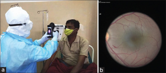
A cross-sectional observational study was conducted on patients affected by COVID-19 who were admitted at our center. Our study included patients with no, mild, and moderate symptoms (nonsevere cases). Intensive care unit (ICU)-admitted patients were excluded considering the difficulty in procuring the fundus image by the handheld camera due to patients positioning. Patients with systemic conditions (diabetes, hypertension, and severe anemia) known to cause retinopathy were also excluded. Bedside anterior segment examination, fundus examination using indirect ophthalmoscopy and fundus imaging of each patient using a nonmydriatic handheld fundus camera was done by a trained ophthalmologist posted for COVID duty.

RESULTS

In a cohort of 138 patients, 94 (68.1%) were men and 44 (31.9%) were women. A total of 276 eyes were evaluated. The mean age of the patients was 38.51 ± 14.4 years. Anterior segment evaluation showed no abnormality in any of the eyes. On fundus screening using nonmydriatic handheld camera, a single streak of superficial retinal hemorrhage was noted at the posterior pole of the fundus in the left eye of one patient (0.72%), which was away from fovea. Laboratory tests revealed low hemoglobin (between 10 and 10.9 g/dL falling under mild Anemia) in 12 patients, elevated total leucocyte count in 6 patients, raised LDH in majority of patients (323 ± 101 Units/L) and elevated CRP (14.6 ± 30.99 mg/L). Rest of the lab parameters were within the normal range.

CONCLUSION

In our study, COVID patients with mild-to-moderate symptoms did not show any inflammatory/infective or vaso-occlusive lesions in the retina attributable to COVID-19 infection, except one patient who had a single streak hemorrhage in the macula away from fovea, probably incidental.

摘要

目的

本研究旨在使用非散瞳手持式眼底相机,研究并记录非重症新型冠状病毒肺炎(COVID-19)阳性患者的视网膜变化。

方法

对我院收治的COVID-19患者进行横断面观察性研究。我们的研究纳入了无症状、轻症和中症患者(非重症病例)。由于手持相机获取眼底图像时患者体位的原因,入住重症监护病房(ICU)的患者被排除。已知会导致视网膜病变的全身性疾病(糖尿病、高血压和重度贫血)患者也被排除。由一名负责COVID相关工作的训练有素的眼科医生对每位患者进行床边眼前节检查、使用间接检眼镜进行眼底检查以及使用非散瞳手持式眼底相机进行眼底成像。

结果

在138例患者队列中,男性94例(68.1%),女性44例(31.9%)。共评估了276只眼。患者的平均年龄为38.51±14.4岁。眼前节评估显示所有眼睛均无异常。使用非散瞳手持式相机进行眼底筛查时,一名患者(0.72%)的左眼眼底后极部发现一条表浅视网膜出血条纹,远离黄斑中心凹。实验室检查显示,12例患者血红蛋白低(10至10.9 g/dL,属于轻度贫血),6例患者白细胞总数升高,大多数患者乳酸脱氢酶升高(323±101单位/L),C反应蛋白升高(14.6±30.99 mg/L)。其余实验室参数均在正常范围内。

结论

在我们的研究中,除一名患者黄斑区远离中心凹处有一条出血条纹(可能为偶然情况)外,轻至中度症状的COVID患者未显示出任何因COVID-19感染导致的视网膜炎症/感染或血管闭塞性病变。

https://cdn.ncbi.nlm.nih.gov/pmc/blobs/3f9a/8186568/e08c14979467/IJO-69-1271-g001.jpg

相似文献

1
Fundus evaluation in COVID-19 positives with non-severe disease.对非重症 COVID-19 阳性患者的眼底评估。
Indian J Ophthalmol. 2021 May;69(5):1271-1274. doi: 10.4103/ijo.IJO_3227_20.
2
Effectiveness and safety of screening for diabetic retinopathy with two nonmydriatic digital images compared with the seven standard stereoscopic photographic fields.与七个标准立体摄影视野相比,用两张非散瞳数字图像筛查糖尿病视网膜病变的有效性和安全性。
Can J Ophthalmol. 2003 Dec;38(7):557-68. doi: 10.1016/s0008-4182(03)80109-6.
3
Diagnostic accuracy of non-mydriatic fundus camera for screening of diabetic retinopathy: A hospital based observational study in Pakistan.非散瞳眼底相机筛查糖尿病视网膜病变的诊断准确性:巴基斯坦一项基于医院的观察性研究。
J Pak Med Assoc. 2019 Mar;69(3):378-382.
4
Image quality and diagnostic accuracy of a handheld nonmydriatic fundus camera: Feasibility of a telemedical approach in screening retinal diseases.手持式免散瞳眼底相机的图像质量和诊断准确性:远程医疗在筛查视网膜疾病中的可行性。
J Chin Med Assoc. 2020 Oct;83(10):962-966. doi: 10.1097/JCMA.0000000000000382.
5
Nonmydriatic ocular fundus photography among headache patients in an emergency department.急诊头痛患者的免散瞳眼底照相。
Neurology. 2013 Jan 29;80(5):432-7. doi: 10.1212/WNL.0b013e31827f0f20. Epub 2013 Jan 2.
6
Diagnostic accuracy and use of nonmydriatic ocular fundus photography by emergency physicians: phase II of the FOTO-ED study.急诊医生使用非散瞳眼底照相的诊断准确性和应用:FOTO-ED 研究第二阶段。
Ann Emerg Med. 2013 Jul;62(1):28-33.e1. doi: 10.1016/j.annemergmed.2013.01.010. Epub 2013 Feb 21.
7
Fundus Lesions in Patients Hospitalized With COVID-19 Infection in Mumbai, India: A Retrospective Review.印度孟买新冠肺炎感染住院患者的眼底病变:一项回顾性研究。
Cureus. 2020 Nov 16;12(11):e11512. doi: 10.7759/cureus.11512.
8
Comparison of two, three and four 45 degrees image fields obtained with the Topcon CRW6 nonmydriatic camera for screening for diabetic retinopathy.使用拓普康CRW6免散瞳相机获得的两个、三个和四个45度图像视野用于糖尿病视网膜病变筛查的比较。
Can J Ophthalmol. 2003 Dec;38(7):569-74. doi: 10.1016/s0008-4182(03)80110-2.
9
The retinal disease screening study: retrospective comparison of nonmydriatic fundus photography and three-dimensional optical coherence tomography for detection of retinal irregularities.视网膜疾病筛查研究:非散瞳眼底照相术与三维光相干断层扫描检测视网膜不规则性的回顾性比较。
Invest Ophthalmol Vis Sci. 2013 Aug 21;54(8):5694-700. doi: 10.1167/iovs.13-12043.
10
Predictors of Photographic Quality with a Handheld Nonmydriatic Fundus Camera Used for Screening of Vision-Threatening Diabetic Retinopathy.用于筛查威胁视力的糖尿病视网膜病变的手持式免散瞳眼底相机的照片质量预测因素
Ophthalmologica. 2017;238(1-2):89-99. doi: 10.1159/000475773. Epub 2017 Jul 5.

引用本文的文献

1
Manifestations of COVID-19 in the posterior eye segment - Up-to-date.新型冠状病毒肺炎在后段眼的表现——最新情况。
Oman J Ophthalmol. 2024 Jun 27;17(2):166-172. doi: 10.4103/ojo.ojo_212_22. eCollection 2024 May-Aug.
2
The prone position in COVID-19 impacts the thickness of peripapillary retinal nerve fiber layers and macular ganglion cell layers.COVID-19 患者采取俯卧位会影响视乳头周围视网膜神经纤维层和黄斑神经节细胞层的厚度。
PLoS One. 2024 May 2;19(5):e0300621. doi: 10.1371/journal.pone.0300621. eCollection 2024.
3
Ocular manifestations of COVID-19: A systematic review of current evidence.

本文引用的文献

1
Ocular findings in patients with coronavirus disease 2019 (COVID-19) in an outbreak hospital.在一家暴发医院中,2019 年冠状病毒病(COVID-19)患者的眼部表现。
J Med Virol. 2021 Feb;93(2):1126-1132. doi: 10.1002/jmv.26412. Epub 2020 Aug 21.
2
COVID-19: Ocular Manifestations and the APAO Prevention Guidelines for Ophthalmic Practices.COVID-19:眼部表现和 APAO 眼科实践预防指南。
Asia Pac J Ophthalmol (Phila). 2020 Jul-Aug;9(4):281-284. doi: 10.1097/APO.0000000000000308.
3
Bilateral anterior uveitis as a part of a multisystem inflammatory syndrome secondary to COVID-19 infection.
新型冠状病毒肺炎的眼部表现:当前证据的系统评价
Prev Med Rep. 2024 Jan 14;38:102608. doi: 10.1016/j.pmedr.2024.102608. eCollection 2024 Feb.
4
The Impact of COVID-19 Infection on Macular Capillary Perfusion.2019冠状病毒病感染对黄斑区毛细血管灌注的影响
Beyoglu Eye J. 2023 Dec 1;8(4):273-279. doi: 10.14744/bej.2023.13549. eCollection 2023.
5
From Bedside to Diagnosis: The Role of Ocular Fundus in Systemic Infections.从床边到诊断:眼底在全身感染中的作用
J Clin Med. 2023 Nov 21;12(23):7216. doi: 10.3390/jcm12237216.
6
The spectrum of COVID-19-associated chorioretinal vasculopathy.新型冠状病毒肺炎相关脉络膜视网膜血管病变的谱系
Am J Ophthalmol Case Rep. 2023 Sep;31:101857. doi: 10.1016/j.ajoc.2023.101857. Epub 2023 May 18.
7
Use of Imaging Technology to Assess the Effect of COVID-19 on Retinal Tissues: A Systematic Review.使用成像技术评估2019冠状病毒病对视网膜组织的影响:一项系统评价
Ophthalmol Ther. 2022 Jun;11(3):1017-1030. doi: 10.1007/s40123-022-00509-8. Epub 2022 Apr 29.
8
Coronavirus Disease 2019-Related Health Disparities in Ophthalmology with a Retrospective Analysis at a Large Academic Public Hospital.大型学术公立医院的回顾性分析:2019年冠状病毒病相关的眼科健康差异
Adv Ophthalmol Optom. 2022 Aug;7(1):311-323. doi: 10.1016/j.yaoo.2022.04.005. Epub 2022 Apr 22.
9
COVID-19-Related Retinal Micro-vasculopathy - A Review of Current Evidence.COVID-19 相关视网膜微血管病变——现有证据的综述。
Am J Ophthalmol. 2022 Mar;235:98-110. doi: 10.1016/j.ajo.2021.09.019. Epub 2021 Sep 26.
双侧前葡萄膜炎作为新型冠状病毒肺炎(COVID-19)感染继发的多系统炎症综合征的一部分。
J Med Virol. 2021 Jan;93(1):139-140. doi: 10.1002/jmv.26229. Epub 2020 Sep 30.
4
Presence of viral RNA of SARS-CoV-2 in conjunctival swab specimens of COVID-19 patients.SARS-CoV-2 的病毒 RNA 存在于 COVID-19 患者的结膜拭子标本中。
Indian J Ophthalmol. 2020 Jun;68(6):1015-1017. doi: 10.4103/ijo.IJO_1287_20.
5
Weathering the COVID-19 storm: Lessons from hematologic cytokine syndromes.应对 COVID-19 风暴:血液细胞因子综合征的经验教训。
Blood Rev. 2021 Jan;45:100707. doi: 10.1016/j.blre.2020.100707. Epub 2020 May 15.
6
COVID-19: progression of disease and intravascular coagulation - present status and future perspectives.COVID-19:疾病进展与血管内凝血——现状与未来展望。
Clin Chem Lab Med. 2020 Jun 25;58(7):1029-1036. doi: 10.1515/cclm-2020-0502.
7
Retinal findings in patients with COVID-19.新型冠状病毒肺炎患者的视网膜病变
Lancet. 2020 May 23;395(10237):1610. doi: 10.1016/S0140-6736(20)31014-X. Epub 2020 May 12.
8
COVID-19 and its implications for thrombosis and anticoagulation.新型冠状病毒肺炎及其对血栓形成和抗凝的影响。
Blood. 2020 Jun 4;135(23):2033-2040. doi: 10.1182/blood.2020006000.
9
Time to consider histologic pattern of lung injury to treat critically ill patients with COVID-19 infection.是时候考虑肺损伤的组织学模式来治疗新冠肺炎感染的重症患者了。
Intensive Care Med. 2020 Jun;46(6):1124-1126. doi: 10.1007/s00134-020-06057-8. Epub 2020 Apr 23.
10
Complex Immune Dysregulation in COVID-19 Patients with Severe Respiratory Failure.COVID-19 患者严重呼吸衰竭的复杂免疫失调。
Cell Host Microbe. 2020 Jun 10;27(6):992-1000.e3. doi: 10.1016/j.chom.2020.04.009. Epub 2020 Apr 21.