Vetsika Eleni-Kyriaki, Sharma Priyanka, Samaras Ioannis, Markou Alexandra, Georgoulias Vassilis, Whiteside Theresa L, Kotsakis Athanasios
School of Medicine, University of Crete, 71003 Heraklion, Greece.
School of Medicine, National and Kapodistrian University of Athens, 11527 Athens, Greece.
Cancers (Basel). 2021 Apr 23;13(9):2041. doi: 10.3390/cancers13092041.
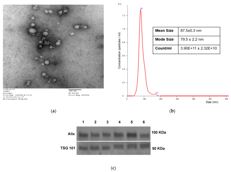
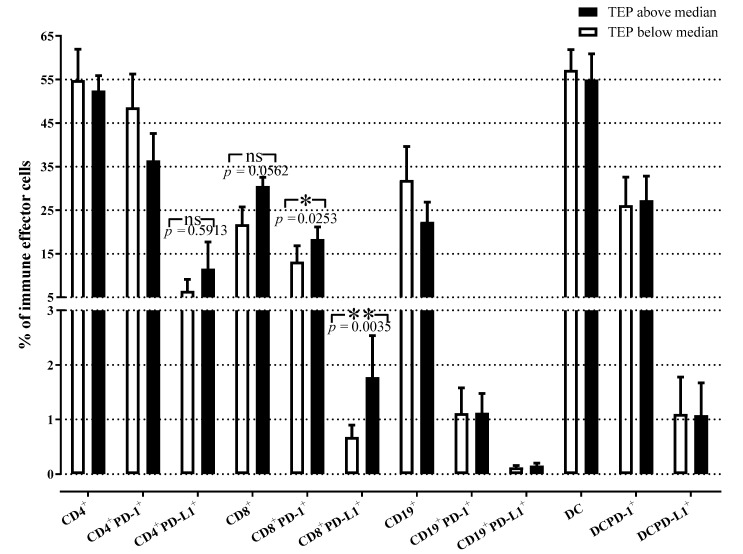
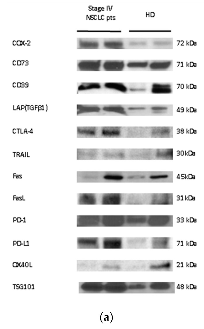
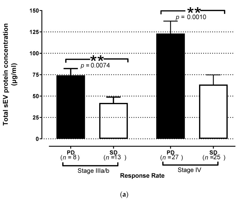
The potential use of plasma-derived small extracellular vesicles (sEV) as predictors of response to therapy and clinical outcome in chemotherapy-naïve patients with non-small-cell lung cancer (NSCLC) was explored. sEV were isolated by size-exclusion chromatography from the plasma of 79 chemotherapy-naïve NSCLC patients and 12 healthy donors (HD). sEV were characterized with regard to protein content, particle size, counts by qNano, morphology by transmission electron microscopy, and molecular profiles by Western blots. PD-1 and PD-L1 expression on circulating immune cells was analysed by flow cytometry. Pre-treatment levels of total sEV protein (TEP) were correlated with overall (OS) and progression-free survival (PFS). The sEV numbers and protein levels were significantly elevated in the plasma of NSCLC patients compared to HD ( = 0.009 and 0.0001, respectively). Baseline TEP levels were higher in patients who developed progressive disease compared to patients with stable disease ( = 0.007 and 0.001, stage III and IV, respectively). Patient-derived sEV were enriched in immunosuppressive proteins as compared to proteins carried by sEV from HD. TEP levels were positively correlated with CD8PD-1 and CD8PD-L1 circulating T cell percentages and were independently associated with poorer PFS ( < 0.00001) and OS ( < 0.00001). Pre-therapy sEV could be useful as non-invasive biomarkers of response to therapy and clinical outcome in NSCLC.
探讨了血浆来源的小细胞外囊泡(sEV)作为初治非小细胞肺癌(NSCLC)患者治疗反应和临床结局预测指标的潜在用途。通过尺寸排阻色谱法从79例初治NSCLC患者和12例健康供体(HD)的血浆中分离出sEV。对sEV的蛋白质含量、颗粒大小、qNano计数、透射电子显微镜形态以及蛋白质印迹法的分子谱进行了表征。通过流式细胞术分析循环免疫细胞上的PD-1和PD-L1表达。将总sEV蛋白(TEP)的治疗前水平与总生存期(OS)和无进展生存期(PFS)进行关联分析。与HD相比,NSCLC患者血浆中的sEV数量和蛋白质水平显著升高(分别为=0.009和0.0001)。与病情稳定的患者相比,疾病进展患者的基线TEP水平更高(分别为=0.007和0.001,III期和IV期)。与HD来源的sEV携带的蛋白质相比,患者来源的sEV富含免疫抑制蛋白。TEP水平与CD8⁺PD-1和CD8⁺PD-L1循环T细胞百分比呈正相关,并且与较差的PFS(<0.00001)和OS(<0.00001)独立相关。治疗前的sEV可作为NSCLC治疗反应和临床结局的非侵入性生物标志物。