• 文献检索
  • 文档翻译
  • 深度研究
  • 学术资讯
  • Suppr Zotero 插件Zotero 插件
  • 邀请有礼
  • 套餐&价格
  • 历史记录
应用&插件
Suppr Zotero 插件Zotero 插件浏览器插件Mac 客户端Windows 客户端微信小程序
定价
高级版会员购买积分包购买API积分包
服务
文献检索文档翻译深度研究API 文档MCP 服务
关于我们
关于 Suppr公司介绍联系我们用户协议隐私条款
关注我们

Suppr 超能文献

核心技术专利:CN118964589B侵权必究
粤ICP备2023148730 号-1Suppr @ 2026

文献检索

告别复杂PubMed语法,用中文像聊天一样搜索,搜遍4000万医学文献。AI智能推荐,让科研检索更轻松。

立即免费搜索

文件翻译

保留排版,准确专业,支持PDF/Word/PPT等文件格式,支持 12+语言互译。

免费翻译文档

深度研究

AI帮你快速写综述,25分钟生成高质量综述,智能提取关键信息,辅助科研写作。

立即免费体验

通过测量脑不同区域的表观扩散系数值来鉴别低级别和高级别胶质瘤肿瘤

Differentiating Between Low- and High-grade Glioma Tumors Measuring Apparent Diffusion Coefficient Values in Various Regions of the Brain.

作者信息

Momeni Farideh, Abedi-Firouzjah Razzagh, Farshidfar Zahra, Taleinezhad Nastaran, Ansari Leila, Razmkon Ali, Banaei Amin, Mehdizadeh Alireza

机构信息

Medical Physics and Biomedical Engineering Department, School of Medicine, Shiraz University of Medical Sciences, Shiraz, Iran.

Research Center for Neuromodulation and Pain, Shiraz University of Medical Sciences, Shiraz, Iran.

出版信息

Oman Med J. 2021 Mar 31;36(2):e251. doi: 10.5001/omj.2021.59. eCollection 2021 Mar.

DOI:10.5001/omj.2021.59
PMID:33936779
原文链接:https://pmc.ncbi.nlm.nih.gov/articles/PMC8077446/
Abstract

OBJECTIVES

Our study aimed to apply the apparent diffusion coefficient (ADC) values to quantify the differences between low- and high-grade glioma tumors.

METHODS

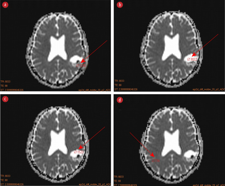
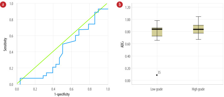
We conducted a multicenter, retrospective study between September to December 2019. Magnetic resonance imaging (MRI) diffusion-weighted images (DWIs), and the pathologic findings of 56 patients with glioma tumors (low grade = 28 and high grade = 28) were assessed to measure the ADC values in the tumor center, tumor edema, boundary area between tumor with normal tissue, and inside the healthy hemisphere. These values were compared between the two groups, and cut-off values were calculated using the receiver operating characteristic curve.

RESULTS

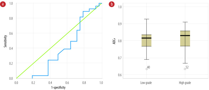
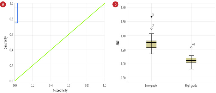
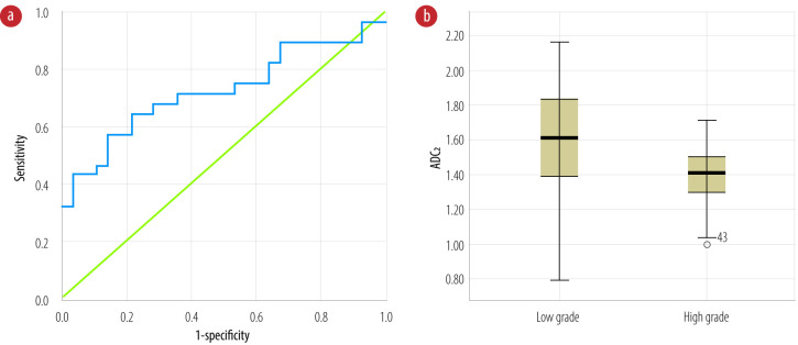
We saw significant differences between the mean ADC values measured in the tumor center and edema between high- and low-grade tumors (< 0.005). The ADC values in the boundary area between tumors with normal tissue and inside healthy hemisphere did not significantly differ in the groups. The ADC values at tumor center and edema were higher than 1.12 × 10 mm/s (sensitivity = 100% and specificity = 96.0%) and 1.15 × 10 mm/s (sensitivity = 75.0% and specificity = 64.0%), respectively, could be classified as low-grade tumors.

CONCLUSIONS

The ADC values from the MRI DWIs in the tumor center and edema could be used as an appropriate method for investigating the differences between low- and high-grade glioma tumors. The ADC values in the boundary area and healthy tissues had no diagnostic values in grading the glioma tumors.

摘要

目的

本研究旨在应用表观扩散系数(ADC)值来量化低级别和高级别胶质瘤肿瘤之间的差异。

方法

我们在2019年9月至12月期间进行了一项多中心回顾性研究。对56例胶质瘤患者(低级别=28例,高级别=28例)的磁共振成像(MRI)扩散加权成像(DWI)及病理结果进行评估,以测量肿瘤中心、肿瘤水肿、肿瘤与正常组织边界区域以及健康脑半球内的ADC值。比较两组之间的这些值,并使用受试者工作特征曲线计算临界值。

结果

我们发现高级别和低级别肿瘤在肿瘤中心和水肿区域测得的平均ADC值之间存在显著差异(<0.005)。肿瘤与正常组织边界区域以及健康脑半球内的ADC值在两组之间无显著差异。肿瘤中心和水肿区域的ADC值分别高于1.12×10⁻³mm²/s(灵敏度=100%,特异度=96.0%)和1.15×10⁻³mm²/s(灵敏度=75.0%,特异度=64.0%)时,可被分类为低级别肿瘤。

结论

MRI DWI在肿瘤中心和水肿区域的ADC值可作为研究低级别和高级别胶质瘤肿瘤差异的合适方法。边界区域和健康组织中的ADC值在胶质瘤肿瘤分级中无诊断价值。

https://cdn.ncbi.nlm.nih.gov/pmc/blobs/9a37/8077446/4a648ad7523a/OMJ-36-02-2000080-f5.jpg
https://cdn.ncbi.nlm.nih.gov/pmc/blobs/9a37/8077446/4278bc756a53/OMJ-36-02-2000080-f1.jpg
https://cdn.ncbi.nlm.nih.gov/pmc/blobs/9a37/8077446/335f88d5041f/OMJ-36-02-2000080-f2.jpg
https://cdn.ncbi.nlm.nih.gov/pmc/blobs/9a37/8077446/0e2190a7e018/OMJ-36-02-2000080-f3.jpg
https://cdn.ncbi.nlm.nih.gov/pmc/blobs/9a37/8077446/9e9f5f3b393b/OMJ-36-02-2000080-f4.jpg
https://cdn.ncbi.nlm.nih.gov/pmc/blobs/9a37/8077446/4a648ad7523a/OMJ-36-02-2000080-f5.jpg

相似文献

1
Differentiating Between Low- and High-grade Glioma Tumors Measuring Apparent Diffusion Coefficient Values in Various Regions of the Brain.通过测量脑不同区域的表观扩散系数值来鉴别低级别和高级别胶质瘤肿瘤
Oman Med J. 2021 Mar 31;36(2):e251. doi: 10.5001/omj.2021.59. eCollection 2021 Mar.
2
Measurements of diagnostic examination performance using quantitative apparent diffusion coefficient and proton MR spectroscopic imaging in the preoperative evaluation of tumor grade in cerebral gliomas.使用定量表观扩散系数和质子磁共振波谱成像对脑胶质瘤术前肿瘤分级进行诊断检查性能的测量。
Eur J Radiol. 2011 Nov;80(2):462-70. doi: 10.1016/j.ejrad.2010.07.017. Epub 2010 Aug 13.
3
Preoperative Grading of Astrocytic Supratentorial Brain Tumors with Diffusion-Weighted Magnetic Resonance Imaging and Apparent Diffusion Coefficient.利用扩散加权磁共振成像和表观扩散系数对幕上星形细胞脑肿瘤进行术前分级
Iran J Radiol. 2016 Feb 17;13(3):e30426. doi: 10.5812/iranjradiol.30426. eCollection 2016 Jul.
4
Quantitative apparent diffusion coefficients in the characterization of brain tumors and associated peritumoral edema.定量表观扩散系数在脑肿瘤及相关瘤周水肿特征描述中的应用
Acta Radiol. 2009 Jul;50(6):682-9. doi: 10.1080/02841850902933123.
5
Diagnostic value of apparent diffusion coefficient in differentiating between high-grade gliomas and brain metastases.表观扩散系数在鉴别高级别胶质瘤与脑转移瘤中的诊断价值
Acta Radiol. 2018 May;59(5):599-605. doi: 10.1177/0284185117727787. Epub 2017 Aug 23.
6
Glioma grading: sensitivity, specificity, positive and negative predictive values of diffusion and perfusion imaging.胶质瘤分级:扩散与灌注成像的敏感性、特异性、阳性和阴性预测值
J Neurooncol. 2009 Aug;94(1):87-96. doi: 10.1007/s11060-009-9807-6. Epub 2009 Feb 20.
7
Voxel-based clustered imaging by multiparameter diffusion tensor images for glioma grading.基于体素的多参数弥散张量成像在胶质瘤分级中的聚类成像。
Neuroimage Clin. 2014 Aug 7;5:396-407. doi: 10.1016/j.nicl.2014.08.001. eCollection 2014.
8
Added Value of Diffusion-Weighted Magnetic Resonance Imaging in Differentiating Musculoskeletal Tumors Using Sensitivity and Specificity: A Retrospective Study and Review of Literature.利用敏感性和特异性评估扩散加权磁共振成像在鉴别肌肉骨骼肿瘤中的附加价值:一项回顾性研究及文献综述
Cureus. 2021 Jan 1;13(1):e12422. doi: 10.7759/cureus.12422.
9
Gliomas: Histogram analysis of apparent diffusion coefficient maps with standard- or high-b-value diffusion-weighted MR imaging--correlation with tumor grade.脑胶质瘤:标准或高 b 值扩散加权磁共振成像表观扩散系数图的直方图分析——与肿瘤分级的相关性。
Radiology. 2011 Dec;261(3):882-90. doi: 10.1148/radiol.11110686. Epub 2011 Oct 3.
10
Preoperative grading of supratentorial gliomas using high or standard b-value diffusion-weighted MR imaging at 3T.使用3T高或标准b值扩散加权磁共振成像对幕上胶质瘤进行术前分级
Diagn Interv Imaging. 2017 Mar;98(3):261-268. doi: 10.1016/j.diii.2016.11.005. Epub 2016 Dec 28.

引用本文的文献

1
Meningioma Prognostic Tool Based on Correlation of Histopathological Grading and Immunohistochemistry.基于组织病理学分级与免疫组化相关性的脑膜瘤预后评估工具
Oman Med J. 2024 Mar 31;39(2):e608. doi: 10.5001/omj.2024.56. eCollection 2024 Mar.
2
Imaging Role in Diagnosis, Prognosis, and Treatment Response Prediction Associated with High-grade Glioma.影像学在高级别胶质瘤的诊断、预后及治疗反应预测中的作用
J Med Signals Sens. 2024 Mar 27;14:7. doi: 10.4103/jmss.jmss_30_22. eCollection 2024.
3
Comparison of the monoexponential and biexponential models of diffusion-weighted magnetic imaging in grading clear-cell renal cell carcinoma: a case-control study.

本文引用的文献

1
Clinical Applications of Arterial Spin Labeling in Brain Tumors.动脉自旋标记在脑肿瘤中的临床应用
J Comput Assist Tomogr. 2019 Jul/Aug;43(4):525-532. doi: 10.1097/RCT.0000000000000873.
2
Differentiating Glioblastomas from Solitary Brain Metastases Using Arterial Spin Labeling Perfusion- and Diffusion Tensor Imaging-Derived Metrics.利用动脉自旋标记灌注和弥散张量成像衍生指标区分胶质母细胞瘤和单发脑转移瘤。
World Neurosurg. 2019 Jul;127:e593-e598. doi: 10.1016/j.wneu.2019.03.213. Epub 2019 Mar 28.
3
Differentiation of Primary Central Nervous System Lymphoma From Glioblastoma: Quantitative Analysis Using Arterial Spin Labeling and Diffusion Tensor Imaging.
扩散加权磁共振成像单指数模型与双指数模型在透明细胞肾细胞癌分级中的比较:一项病例对照研究
Transl Androl Urol. 2024 May 31;13(5):792-801. doi: 10.21037/tau-24-141. Epub 2024 May 28.
4
Frailty in patients with IDH-mutant gliomas: experience from a high-volume tumor center.IDH 突变型脑胶质瘤患者的虚弱状况:来自大容量肿瘤中心的经验。
J Neurooncol. 2024 Jul;168(3):435-443. doi: 10.1007/s11060-024-04685-4. Epub 2024 Jun 4.
5
Prognostic Value of Apparent Diffusion Coefficient (ADC) in Patients with Diffuse Gliomas.表观扩散系数(ADC)在弥漫性胶质瘤患者中的预后价值
Cancers (Basel). 2024 Feb 6;16(4):681. doi: 10.3390/cancers16040681.
6
Predicting the WHO Grading of Pediatric Brain Tumors Based on Their MRI Appearance: A Retrospective Study.基于MRI表现预测儿童脑肿瘤的世界卫生组织分级:一项回顾性研究。
Cureus. 2023 Oct 19;15(10):e47333. doi: 10.7759/cureus.47333. eCollection 2023 Oct.
原发性中枢神经系统淋巴瘤与胶质母细胞瘤的鉴别:使用动脉自旋标记和扩散张量成像的定量分析
World Neurosurg. 2019 Mar;123:e303-e309. doi: 10.1016/j.wneu.2018.11.155. Epub 2018 Nov 29.
4
Correlation between apparent diffusion coefficient (ADC) and cellularity is different in several tumors: a meta-analysis.表观扩散系数(ADC)与细胞密度在几种肿瘤中的相关性不同:一项荟萃分析。
Oncotarget. 2017 May 10;8(35):59492-59499. doi: 10.18632/oncotarget.17752. eCollection 2017 Aug 29.
5
Diagnostic value of apparent diffusion coefficient in differentiating between high-grade gliomas and brain metastases.表观扩散系数在鉴别高级别胶质瘤与脑转移瘤中的诊断价值
Acta Radiol. 2018 May;59(5):599-605. doi: 10.1177/0284185117727787. Epub 2017 Aug 23.
6
Using Nanoparticles in Medicine for Liver Cancer Imaging.纳米颗粒在肝癌成像医学中的应用。
Oman Med J. 2017 Jul;32(4):269-274. doi: 10.5001/omj.2017.54.
7
Differentiation of Glioblastoma from Brain Metastasis: Qualitative and Quantitative Analysis Using Arterial Spin Labeling MR Imaging.胶质母细胞瘤与脑转移瘤的鉴别:使用动脉自旋标记磁共振成像的定性和定量分析
PLoS One. 2016 Nov 18;11(11):e0166662. doi: 10.1371/journal.pone.0166662. eCollection 2016.
8
Assessment of diffusion tensor imaging metrics in differentiating low-grade from high-grade gliomas.利用扩散张量成像指标鉴别低级别与高级别胶质瘤的研究
Neuroradiol J. 2016 Oct;29(5):400-7. doi: 10.1177/1971400916665382. Epub 2016 Aug 25.
9
Automatic Analysis of Cellularity in Glioblastoma and Correlation with ADC Using Trajectory Analysis and Automatic Nuclei Counting.使用轨迹分析和自动细胞核计数对胶质母细胞瘤中的细胞密度进行自动分析及其与表观扩散系数的相关性
PLoS One. 2016 Jul 28;11(7):e0160250. doi: 10.1371/journal.pone.0160250. eCollection 2016.
10
Radiological indeterminate vestibular schwannoma and meningioma in cerebellopontine angle area: differentiating using whole-tumor histogram analysis of apparent diffusion coefficient.桥小脑角区放射学表现不典型的前庭神经鞘瘤和脑膜瘤:利用表观扩散系数的全肿瘤直方图分析进行鉴别
Int J Neurosci. 2017 Feb;127(2):183-190. doi: 10.3109/00207454.2016.1164157. Epub 2016 Apr 6.