Anam Choirul, Arif Idam, Haryanto Freddy, Lestari Fauzia P, Widita Rena, Budi Wahyu S, Sutanto Heri, Adi Kusworo, Fujibuchi Toshioh, Dougherty Geoff
PhD, Department of Physics, Faculty of Sciences and Mathematics, Diponegoro University, Jl. Prof. Soedarto SH, Tembalang, Semarang 50275, Central Java, Indonesia.
PhD, Department of Physics, Faculty of Mathematics and Natural Sciences, Bandung Institute of Technology, Ganesha 10, Bandung 40132, West Java, Indonesia.
J Biomed Phys Eng. 2021 Apr 1;11(2):163-174. doi: 10.31661/jbpe.v0i0.1198. eCollection 2021 Apr.
It is necessary to have an automated noise measurement system working accurately to optimize dose in computerized tomography (CT) examinations.
This study aims to develop an algorithm to automate noise measurement that can be implemented in CT images of all body regions.
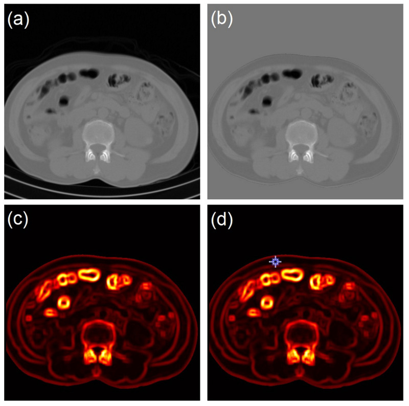
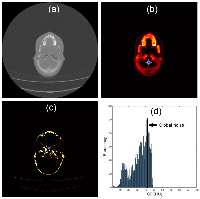
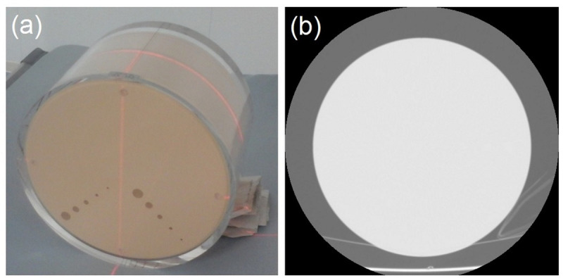
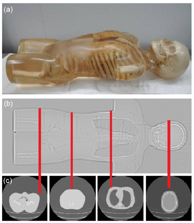
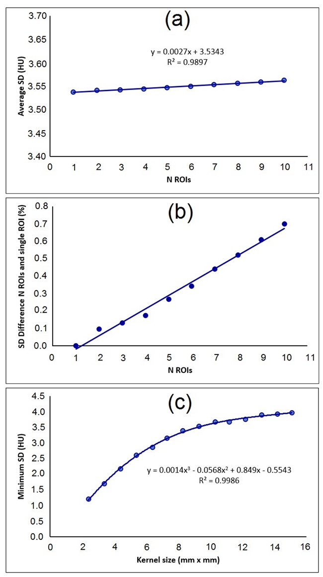
In this retrospective study, our automated noise measurement method consists of three steps as follows: the first is segmenting the image of the patient. The second is developing a standard deviation (SD) map by calculating the SD value for each pixel with a sliding window operation. The third step is estimating the noise as the smallest SD from the SD map. The proposed method was applied to the images of a homogenous phantom and a full body adult anthropomorphic phantom, and retrospectively applied to 27 abdominal images of patients.
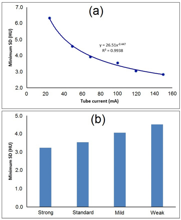
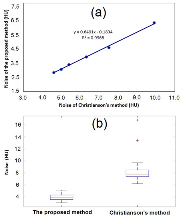
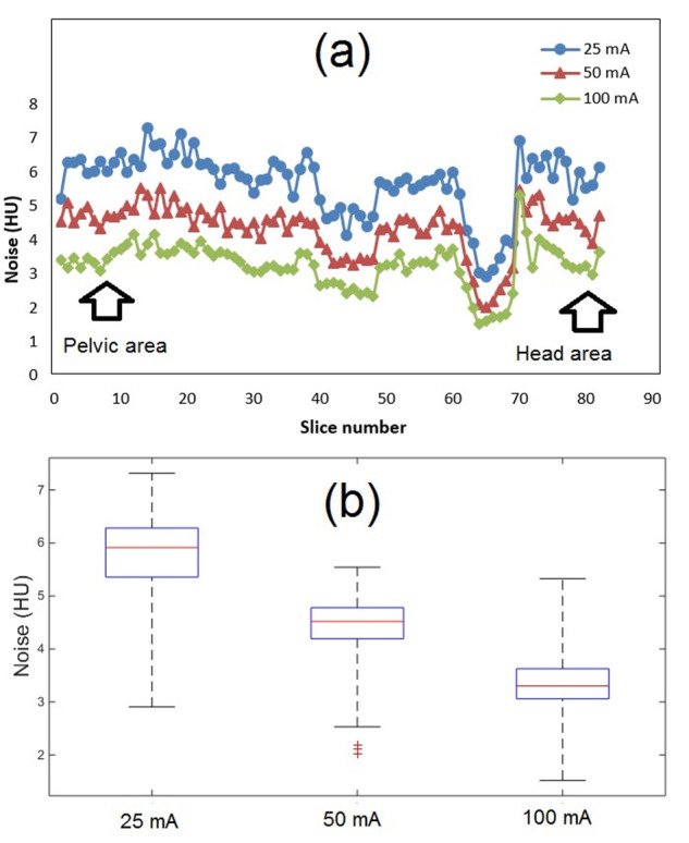
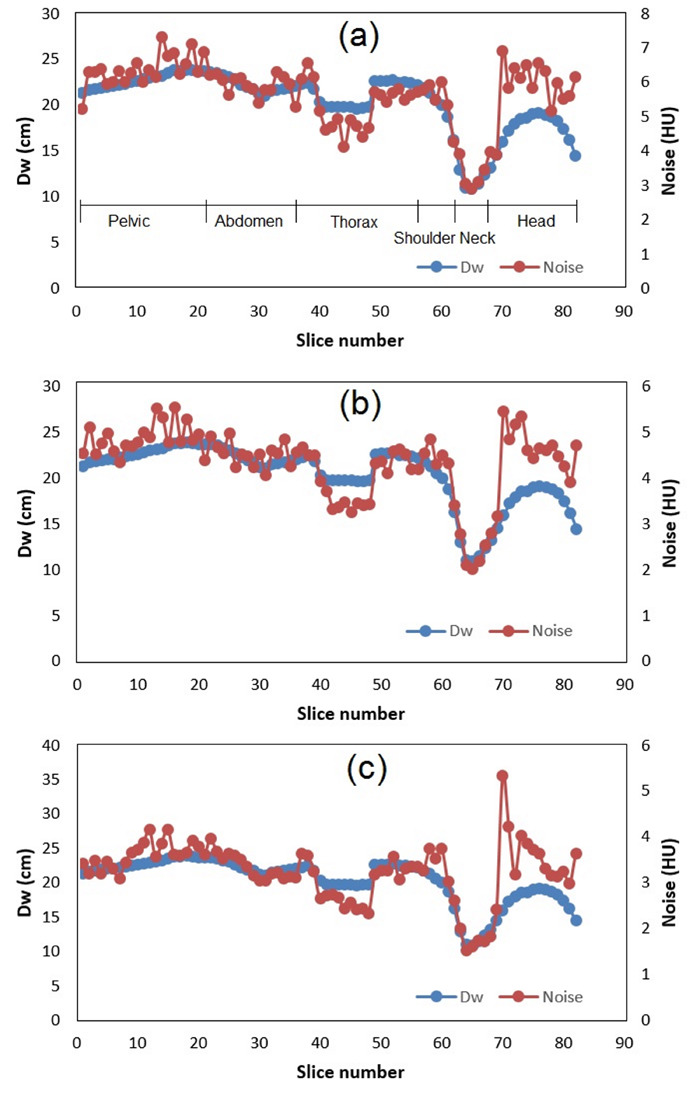
For a homogeneous phantom, the noises calculated using our proposed and previous algorithms have a linear correlation with R = 0.997. It is found that the noise magnitude closely follows the magnitude of the water equivalent diameter (D) in all body regions. The proposed algorithm is able to distinguish the noise magnitude due to variations in tube currents and different noise suppression techniques such as strong, standard, mild, and weak ones in a reconstructed image using the AIDR 3D algorithm.
An automated noise calculation has been proposed and successfully implemented in all body regions. It is not only accurate and easy to implement but also not influenced by the subjectivity of user.
需要一个准确运行的自动噪声测量系统来优化计算机断层扫描(CT)检查中的剂量。
本研究旨在开发一种可在所有身体部位的CT图像中实现的自动噪声测量算法。
在这项回顾性研究中,我们的自动噪声测量方法包括以下三个步骤:第一步是分割患者图像。第二步是通过使用滑动窗口操作计算每个像素的标准差(SD)值来生成标准差图。第三步是将噪声估计为标准差图中的最小标准差。所提出的方法应用于均匀体模和全身成人仿真体模的图像,并回顾性应用于27例患者的腹部图像。
对于均匀体模,使用我们提出的算法和先前算法计算出的噪声具有线性相关性,R = 0.997。发现在所有身体部位,噪声大小与水等效直径(D)的大小密切相关。所提出的算法能够在使用AIDR 3D算法重建的图像中,根据管电流的变化以及不同的噪声抑制技术(如强、标准、弱和极弱)来区分噪声大小。
已提出一种自动噪声计算方法,并在所有身体部位成功实现。它不仅准确且易于实现,而且不受用户主观性的影响。