• 文献检索
  • 文档翻译
  • 深度研究
  • 学术资讯
  • Suppr Zotero 插件Zotero 插件
  • 邀请有礼
  • 套餐&价格
  • 历史记录
应用&插件
Suppr Zotero 插件Zotero 插件浏览器插件Mac 客户端Windows 客户端微信小程序
定价
高级版会员购买积分包购买API积分包
服务
文献检索文档翻译深度研究API 文档MCP 服务
关于我们
关于 Suppr公司介绍联系我们用户协议隐私条款
关注我们

Suppr 超能文献

核心技术专利:CN118964589B侵权必究
粤ICP备2023148730 号-1Suppr @ 2026

文献检索

告别复杂PubMed语法,用中文像聊天一样搜索,搜遍4000万医学文献。AI智能推荐,让科研检索更轻松。

立即免费搜索

文件翻译

保留排版,准确专业,支持PDF/Word/PPT等文件格式,支持 12+语言互译。

免费翻译文档

深度研究

AI帮你快速写综述,25分钟生成高质量综述,智能提取关键信息,辅助科研写作。

立即免费体验

通过对比增强乳腺钼靶摄影提取纹理特征的放射组学和人工智能分析在乳腺病变分类中的应用

Radiomics and Artificial Intelligence Analysis with Textural Metrics Extracted by Contrast-Enhanced Mammography in the Breast Lesions Classification.

作者信息

Fusco Roberta, Piccirillo Adele, Sansone Mario, Granata Vincenza, Rubulotta Maria Rosaria, Petrosino Teresa, Barretta Maria Luisa, Vallone Paolo, Di Giacomo Raimondo, Esposito Emanuela, Di Bonito Maurizio, Petrillo Antonella

机构信息

Radiology Division, Istituto Nazionale Tumori-IRCCS-Fondazione G. Pascale, 80131 Naples, Italy.

Department of Electrical Engineering and Information Technologies, Università degli Studi di Napoli Federico II, 80125 Naples, Italy.

出版信息

Diagnostics (Basel). 2021 Apr 30;11(5):815. doi: 10.3390/diagnostics11050815.

DOI:10.3390/diagnostics11050815
PMID:33946333
原文链接:https://pmc.ncbi.nlm.nih.gov/articles/PMC8146084/
Abstract

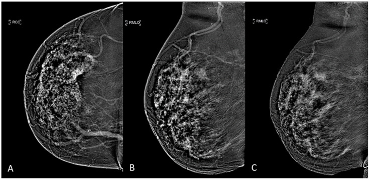
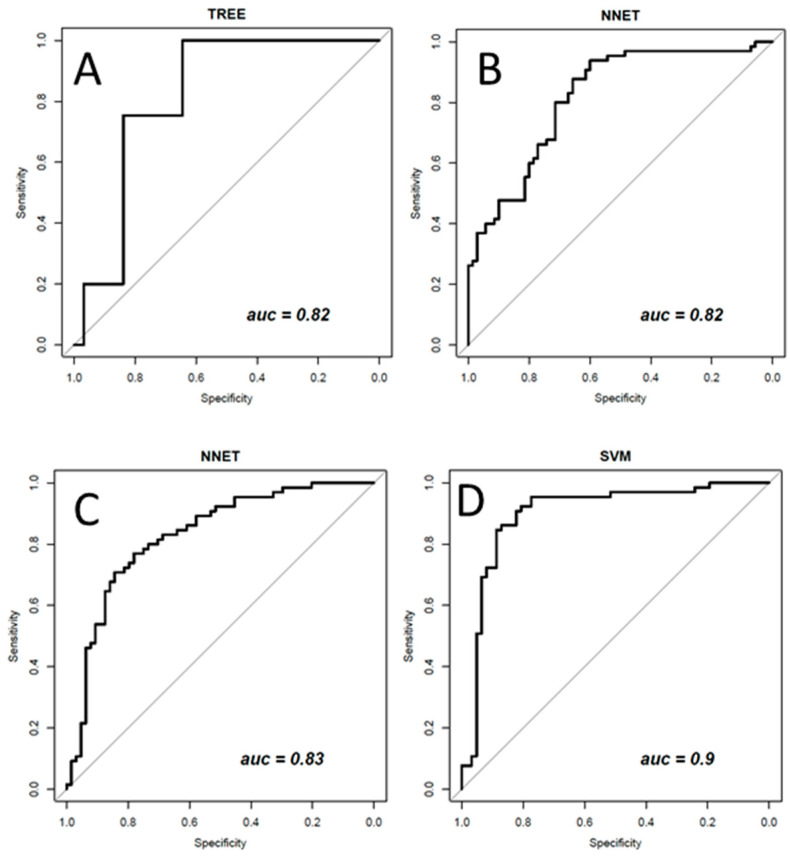
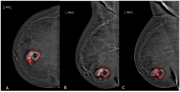
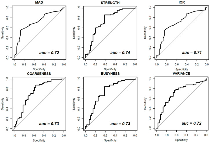
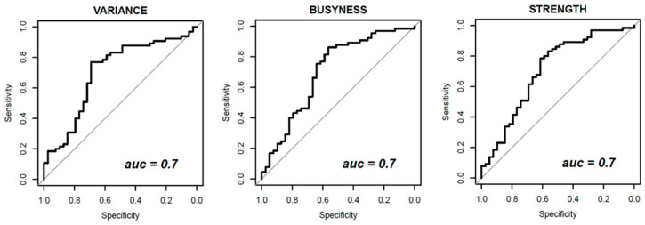
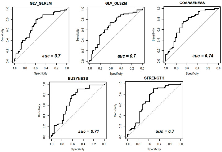
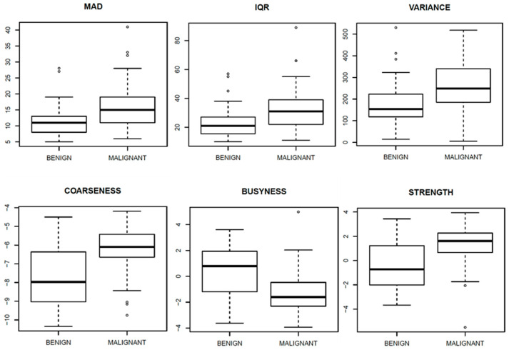
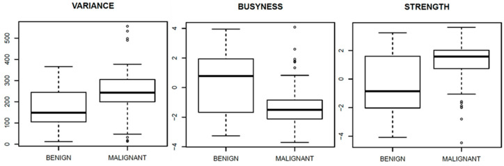
The aim of the study was to estimate the diagnostic accuracy of textural features extracted by dual-energy contrast-enhanced mammography (CEM) images, by carrying out univariate and multivariate statistical analyses including artificial intelligence approaches. In total, 80 patients with known breast lesion were enrolled in this prospective study according to regulations issued by the local Institutional Review Board. All patients underwent dual-energy CEM examination in both craniocaudally (CC) and double acquisition of mediolateral oblique (MLO) projections (early and late). The reference standard was pathology from a surgical specimen for malignant lesions and pathology from a surgical specimen or fine needle aspiration cytology, core or Tru-Cut needle biopsy, and vacuum assisted breast biopsy for benign lesions. In total, 104 samples of 80 patients were analyzed. Furthermore, 48 textural parameters were extracted by manually segmenting regions of interest. Univariate and multivariate approaches were performed: non-parametric Wilcoxon-Mann-Whitney test; receiver operating characteristic (ROC), linear classifier (LDA), decision tree (DT), k-nearest neighbors (KNN), artificial neural network (NNET), and support vector machine (SVM) were utilized. A balancing approach and feature selection methods were used. The univariate analysis showed low accuracy and area under the curve (AUC) for all considered features. Instead, in the multivariate textural analysis, the best performance considering the CC view (accuracy (ACC) = 0.75; AUC = 0.82) was reached with a DT trained with leave-one-out cross-variation (LOOCV) and balanced data (with adaptive synthetic (ADASYN) function) and a subset of three robust textural features (MAD, VARIANCE, and LRLGE). The best performance (ACC = 0.77; AUC = 0.83) considering the early-MLO view was reached with a NNET trained with LOOCV and balanced data (with ADASYN function) and a subset of ten robust features (MEAN, MAD, RANGE, IQR, VARIANCE, CORRELATION, RLV, COARSNESS, BUSYNESS, and STRENGTH). The best performance (ACC = 0.73; AUC = 0.82) considering the late-MLO view was reached with a NNET trained with LOOCV and balanced data (with ADASYN function) and a subset of eleven robust features (MODE, MEDIAN, RANGE, RLN, LRLGE, RLV, LZLGE, GLV_GLSZM, ZSV, COARSNESS, and BUSYNESS). Multivariate analyses using pattern recognition approaches, considering 144 textural features extracted from all three mammographic projections (CC, early MLO, and late MLO), optimized by adaptive synthetic sampling and feature selection operations obtained the best results (ACC = 0.87; AUC = 0.90) and showed the best performance in the discrimination of benign and malignant lesions.

摘要

本研究的目的是通过进行包括人工智能方法在内的单变量和多变量统计分析,评估双能对比增强乳腺摄影(CEM)图像提取的纹理特征的诊断准确性。根据当地机构审查委员会发布的规定,本前瞻性研究共纳入了80例已知乳腺病变的患者。所有患者均在头尾位(CC)以及内外斜位(MLO)投影的双期采集(早期和晚期)下接受了双能CEM检查。恶性病变的参考标准是手术标本的病理结果,良性病变的参考标准是手术标本或细针穿刺细胞学检查、粗针或Tru-Cut针活检以及真空辅助乳腺活检的病理结果。总共对80例患者的104个样本进行了分析。此外,通过手动分割感兴趣区域提取了48个纹理参数。进行了单变量和多变量分析方法:非参数Wilcoxon-Mann-Whitney检验;使用了受试者操作特征(ROC)、线性分类器(LDA)、决策树(DT)、k近邻(KNN)、人工神经网络(NNET)和支持向量机(SVM)。采用了平衡方法和特征选择方法。单变量分析显示,所有考虑的特征的准确性和曲线下面积(AUC)都较低。相反,在多变量纹理分析中,考虑CC视图时(准确率(ACC)=0.75;AUC=0.82),使用留一法交叉验证(LOOCV)和平衡数据(使用自适应合成(ADASYN)函数)训练的DT以及三个稳健纹理特征(平均绝对偏差(MAD)、方差(VARIANCE)和长行程游程长度矩阵(LRLGE))的子集达到了最佳性能。考虑早期MLO视图时(ACC=0.77;AUC=0.83),使用LOOCV和平衡数据(使用ADASYN函数)训练的NNET以及十个稳健特征(均值(MEAN)、MAD、范围(RANGE)、四分位距(IQR)、方差、相关性(CORRELATION)、游程长度百分比(RLV)、粗糙度(COARSNESS)、繁忙度(BUSYNESS)和强度(STRENGTH))的子集达到了最佳性能。考虑晚期MLO视图时(ACC=0.73;AUC=0.82),使用LOOCV和平衡数据(使用ADASYN函数)训练的NNET以及十一个稳健特征(众数(MODE)、中位数(MEDIAN)、RANGE、游程长度非零(RLN)、LRLGE、RLV、短行程游程长度矩阵(LZLGE)、灰度共生矩阵(GLV_GLSZM)、零阶矩(ZSV)、COARSNESS和BUSYNESS)的子集达到了最佳性能。使用模式识别方法进行多变量分析,考虑从所有三个乳腺摄影投影(CC、早期MLO和晚期MLO)中提取的144个纹理特征,通过自适应合成采样和特征选择操作进行优化,获得了最佳结果(ACC=0.87;AUC=0.90),并且在区分良性和恶性病变方面表现出最佳性能。

https://cdn.ncbi.nlm.nih.gov/pmc/blobs/25a3/8146084/be72479d9825/diagnostics-11-00815-g009.jpg
https://cdn.ncbi.nlm.nih.gov/pmc/blobs/25a3/8146084/fe65feb67b32/diagnostics-11-00815-g001.jpg
https://cdn.ncbi.nlm.nih.gov/pmc/blobs/25a3/8146084/0cdab036f195/diagnostics-11-00815-g002.jpg
https://cdn.ncbi.nlm.nih.gov/pmc/blobs/25a3/8146084/622f068c114a/diagnostics-11-00815-g003.jpg
https://cdn.ncbi.nlm.nih.gov/pmc/blobs/25a3/8146084/9d88f2c93e1a/diagnostics-11-00815-g004.jpg
https://cdn.ncbi.nlm.nih.gov/pmc/blobs/25a3/8146084/ccbf391e2708/diagnostics-11-00815-g005.jpg
https://cdn.ncbi.nlm.nih.gov/pmc/blobs/25a3/8146084/f165a0265bf0/diagnostics-11-00815-g006.jpg
https://cdn.ncbi.nlm.nih.gov/pmc/blobs/25a3/8146084/90858ff4a8d2/diagnostics-11-00815-g007.jpg
https://cdn.ncbi.nlm.nih.gov/pmc/blobs/25a3/8146084/9e583f6d082e/diagnostics-11-00815-g008.jpg
https://cdn.ncbi.nlm.nih.gov/pmc/blobs/25a3/8146084/be72479d9825/diagnostics-11-00815-g009.jpg

相似文献

1
Radiomics and Artificial Intelligence Analysis with Textural Metrics Extracted by Contrast-Enhanced Mammography in the Breast Lesions Classification.通过对比增强乳腺钼靶摄影提取纹理特征的放射组学和人工智能分析在乳腺病变分类中的应用
Diagnostics (Basel). 2021 Apr 30;11(5):815. doi: 10.3390/diagnostics11050815.
2
Prediction of Breast Cancer Histological Outcome by Radiomics and Artificial Intelligence Analysis in Contrast-Enhanced Mammography.在对比增强乳腺钼靶摄影中通过影像组学和人工智能分析预测乳腺癌组织学结果
Cancers (Basel). 2022 Apr 25;14(9):2132. doi: 10.3390/cancers14092132.
3
Radiomic and Artificial Intelligence Analysis with Textural Metrics Extracted by Contrast-Enhanced Mammography and Dynamic Contrast Magnetic Resonance Imaging to Detect Breast Malignant Lesions.基于对比增强乳腺摄影和动态对比磁共振成像提取纹理特征的放射组学和人工智能分析在检测乳腺恶性病变中的应用。
Curr Oncol. 2022 Mar 13;29(3):1947-1966. doi: 10.3390/curroncol29030159.
4
A multicentric study of radiomics and artificial intelligence analysis on contrast-enhanced mammography to identify different histotypes of breast cancer.多中心研究:基于对比增强乳腺摄影的影像组学和人工智能分析,以识别不同乳腺癌组织学分型。
Radiol Med. 2024 Jun;129(6):864-878. doi: 10.1007/s11547-024-01817-8. Epub 2024 May 17.
5
EOB-MR Based Radiomics Analysis to Assess Clinical Outcomes following Liver Resection in Colorectal Liver Metastases.基于增强外泌体磁共振成像的放射组学分析评估结直肠癌肝转移肝切除术后的临床结局
Cancers (Basel). 2022 Feb 27;14(5):1239. doi: 10.3390/cancers14051239.
6
Contrast MR-Based Radiomics and Machine Learning Analysis to Assess Clinical Outcomes following Liver Resection in Colorectal Liver Metastases: A Preliminary Study.基于对比增强磁共振成像的影像组学与机器学习分析评估结直肠癌肝转移肝切除术后的临床结局:一项初步研究
Cancers (Basel). 2022 Feb 22;14(5):1110. doi: 10.3390/cancers14051110.
7
Radiomics textural features by MR imaging to assess clinical outcomes following liver resection in colorectal liver metastases.通过磁共振成像的影像组学纹理特征评估结直肠癌肝转移肝切除术后的临床结局。
Radiol Med. 2022 May;127(5):461-470. doi: 10.1007/s11547-022-01477-6. Epub 2022 Mar 26.
8
Mammographic masses characterization based on localized texture and dataset fractal analysis using linear, neural and support vector machine classifiers.基于局部纹理和数据集分形分析,使用线性、神经网络和支持向量机分类器对乳腺X线摄影肿块进行特征描述。
Artif Intell Med. 2006 Jun;37(2):145-62. doi: 10.1016/j.artmed.2006.03.002. Epub 2006 May 23.
9
A multi-stage fusion framework to classify breast lesions using deep learning and radiomics features computed from four-view mammograms.一种基于深度学习和从四视图乳房 X 光片中计算出的放射组学特征的多阶段融合框架,用于对乳腺病变进行分类。
Med Phys. 2023 Dec;50(12):7670-7683. doi: 10.1002/mp.16419. Epub 2023 Apr 21.
10
CT-Based Radiomics Analysis to Predict Histopathological Outcomes Following Liver Resection in Colorectal Liver Metastases.基于CT的影像组学分析预测结直肠癌肝转移肝切除术后的组织病理学结果
Cancers (Basel). 2022 Mar 24;14(7):1648. doi: 10.3390/cancers14071648.

引用本文的文献

1
Comparative Evaluation of Machine Learning-Based Radiomics and Deep Learning for Breast Lesion Classification in Mammography.基于机器学习的放射组学和深度学习在乳腺钼靶摄影中对乳腺病变分类的比较评估
Diagnostics (Basel). 2025 Apr 9;15(8):953. doi: 10.3390/diagnostics15080953.
2
Diagnostic test accuracy of AI-assisted mammography for breast imaging: a narrative review.人工智能辅助乳腺钼靶摄影在乳腺成像中的诊断测试准确性:一项叙述性综述。
PeerJ Comput Sci. 2025 Feb 19;11:e2476. doi: 10.7717/peerj-cs.2476. eCollection 2025.
3
Predicting axillary lymph node metastasis in breast cancer using a multimodal radiomics and deep learning model.

本文引用的文献

1
Radiomic Analysis in Contrast-Enhanced Spectral Mammography for Predicting Breast Cancer Histological Outcome.对比增强光谱乳腺摄影术的放射组学分析用于预测乳腺癌组织学结果
Diagnostics (Basel). 2020 Sep 17;10(9):708. doi: 10.3390/diagnostics10090708.
2
The Image Biomarker Standardization Initiative: Standardized Quantitative Radiomics for High-Throughput Image-based Phenotyping.影像生物标志物标准化倡议:高通量基于影像表型的标准化定量放射组学。
Radiology. 2020 May;295(2):328-338. doi: 10.1148/radiol.2020191145. Epub 2020 Mar 10.
3
Invasive ductal breast cancer: preoperative predict Ki-67 index based on radiomics of ADC maps.
使用多模态放射组学和深度学习模型预测乳腺癌腋窝淋巴结转移
Front Immunol. 2024 Dec 13;15:1482020. doi: 10.3389/fimmu.2024.1482020. eCollection 2024.
4
An updated overview of radiomics-based artificial intelligence (AI) methods in breast cancer screening and diagnosis.基于放射组学的人工智能(AI)方法在乳腺癌筛查和诊断中的最新综述。
Radiol Phys Technol. 2024 Dec;17(4):795-818. doi: 10.1007/s12194-024-00842-6. Epub 2024 Sep 16.
5
Radiomics analysis of intratumoral and different peritumoral regions from multiparametric MRI for evaluating HER2 status of breast cancer: A comparative study.基于多参数磁共振成像对乳腺癌瘤内及不同瘤周区域进行影像组学分析以评估HER2状态:一项对比研究
Heliyon. 2024 Apr 4;10(7):e28722. doi: 10.1016/j.heliyon.2024.e28722. eCollection 2024 Apr 15.
6
Empowering breast cancer diagnosis and radiology practice: advances in artificial intelligence for contrast-enhanced mammography.增强乳腺癌诊断与放射学实践:人工智能在乳腺增强钼靶成像中的进展
Front Radiol. 2024 Jan 5;3:1326831. doi: 10.3389/fradi.2023.1326831. eCollection 2023.
7
A Statistical Approach to Assess the Robustness of Radiomics Features in the Discrimination of Mammographic Lesions.一种评估乳腺钼靶病变鉴别中影像组学特征稳健性的统计方法。
J Pers Med. 2023 Jul 7;13(7):1104. doi: 10.3390/jpm13071104.
8
Freehand 1.5T MR-Guided Vacuum-Assisted Breast Biopsy (MR-VABB): Contribution of Radiomics to the Differentiation of Benign and Malignant Lesions.徒手1.5T磁共振引导下真空辅助乳腺活检(MR-VABB):影像组学对乳腺良恶性病变鉴别的贡献
Diagnostics (Basel). 2023 Mar 7;13(6):1007. doi: 10.3390/diagnostics13061007.
9
The utilization of artificial intelligence applications to improve breast cancer detection and prognosis.利用人工智能应用提高乳腺癌的检测和预后。
Saudi Med J. 2023 Feb;44(2):119-127. doi: 10.15537/smj.2023.44.2.20220611.
10
Machine Learning Approaches with Textural Features to Calculate Breast Density on Mammography.基于纹理特征的机器学习方法计算乳腺 X 线摄影中的乳腺密度。
Curr Oncol. 2023 Jan 7;30(1):839-853. doi: 10.3390/curroncol30010064.
浸润性导管乳腺癌:基于 ADC 图纹理分析的术前预测 Ki-67 指数。
Radiol Med. 2020 Feb;125(2):109-116. doi: 10.1007/s11547-019-01100-1. Epub 2019 Nov 6.
4
Contrast-Enhanced Mammography and Radiomics Analysis for Noninvasive Breast Cancer Characterization: Initial Results.对比增强乳腺摄影和放射组学分析在无创性乳腺癌特征描述中的应用:初步结果。
Mol Imaging Biol. 2020 Jun;22(3):780-787. doi: 10.1007/s11307-019-01423-5.
5
Fully Automated Support System for Diagnosis of Breast Cancer in Contrast-Enhanced Spectral Mammography Images.用于对比增强光谱乳腺钼靶图像中乳腺癌诊断的全自动支持系统
J Clin Med. 2019 Jun 21;8(6):891. doi: 10.3390/jcm8060891.
6
A New Challenge for Radiologists: Radiomics in Breast Cancer.放射科医生面临的新挑战:乳腺癌的影像组学。
Biomed Res Int. 2018 Oct 8;2018:6120703. doi: 10.1155/2018/6120703. eCollection 2018.
7
Contrast Enhanced Spectral Mammography: A Review.对比增强光谱乳腺摄影术:综述
Semin Ultrasound CT MR. 2018 Feb;39(1):70-79. doi: 10.1053/j.sult.2017.08.005. Epub 2017 Aug 24.
8
Digital breast tomosynthesis (DBT): recommendations from the Italian College of Breast Radiologists (ICBR) by the Italian Society of Medical Radiology (SIRM) and the Italian Group for Mammography Screening (GISMa).数字乳腺断层合成(DBT):意大利医学放射学会(SIRM)和意大利乳腺钼靶筛查小组(GISMa)下属的意大利乳腺放射科医师学会(ICBR)发布的建议
Radiol Med. 2017 Oct;122(10):723-730. doi: 10.1007/s11547-017-0769-z. Epub 2017 May 25.
9
A Novel Ensemble Method for Imbalanced Data Learning: Bagging of Extrapolation-SMOTE SVM.一种用于不平衡数据学习的新型集成方法:外推-SMOTE支持向量机的装袋法
Comput Intell Neurosci. 2017;2017:1827016. doi: 10.1155/2017/1827016. Epub 2017 Jan 30.
10
Contrast-enhanced spectral mammography (CESM) versus breast magnetic resonance imaging (MRI): A retrospective comparison in 66 breast lesions.对比增强光谱乳腺摄影(CESM)与乳腺磁共振成像(MRI):66例乳腺病变的回顾性比较
Diagn Interv Imaging. 2017 Feb;98(2):113-123. doi: 10.1016/j.diii.2016.08.013. Epub 2016 Sep 26.