• 文献检索
  • 文档翻译
  • 深度研究
  • 学术资讯
  • Suppr Zotero 插件Zotero 插件
  • 邀请有礼
  • 套餐&价格
  • 历史记录
应用&插件
Suppr Zotero 插件Zotero 插件浏览器插件Mac 客户端Windows 客户端微信小程序
定价
高级版会员购买积分包购买API积分包
服务
文献检索文档翻译深度研究API 文档MCP 服务
关于我们
关于 Suppr公司介绍联系我们用户协议隐私条款
关注我们

Suppr 超能文献

核心技术专利:CN118964589B侵权必究
粤ICP备2023148730 号-1Suppr @ 2026

文献检索

告别复杂PubMed语法,用中文像聊天一样搜索,搜遍4000万医学文献。AI智能推荐,让科研检索更轻松。

立即免费搜索

文件翻译

保留排版,准确专业,支持PDF/Word/PPT等文件格式,支持 12+语言互译。

免费翻译文档

深度研究

AI帮你快速写综述,25分钟生成高质量综述,智能提取关键信息,辅助科研写作。

立即免费体验

基于增强外泌体磁共振成像的放射组学分析评估结直肠癌肝转移肝切除术后的临床结局

EOB-MR Based Radiomics Analysis to Assess Clinical Outcomes following Liver Resection in Colorectal Liver Metastases.

作者信息

Granata Vincenza, Fusco Roberta, De Muzio Federica, Cutolo Carmen, Setola Sergio Venanzio, Dell'Aversana Federica, Ottaiano Alessandro, Nasti Guglielmo, Grassi Roberta, Pilone Vincenzo, Miele Vittorio, Brunese Maria Chiara, Tatangelo Fabiana, Izzo Francesco, Petrillo Antonella

机构信息

Division of Radiology, Istituto Nazionale Tumori IRCCS Fondazione Pascale-IRCCS di Napoli, 80131 Napoli, Italy.

Medical Oncology Division, Igea SpA, 41012 Carpi, Italy.

出版信息

Cancers (Basel). 2022 Feb 27;14(5):1239. doi: 10.3390/cancers14051239.

DOI:10.3390/cancers14051239
PMID:35267544
原文链接:https://pmc.ncbi.nlm.nih.gov/articles/PMC8909637/
Abstract

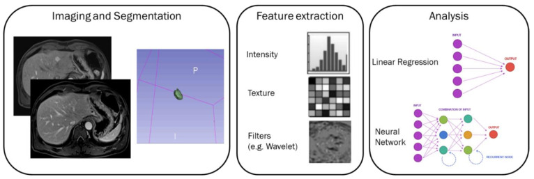
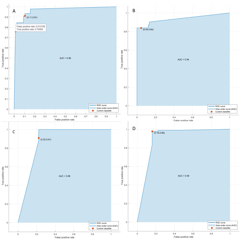
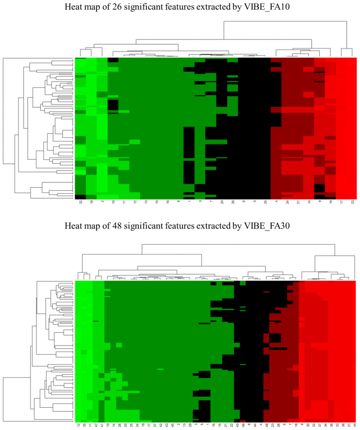
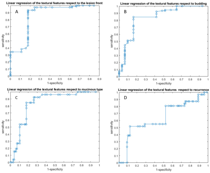
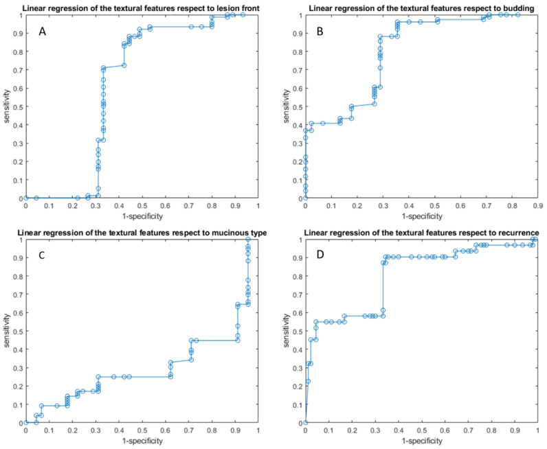
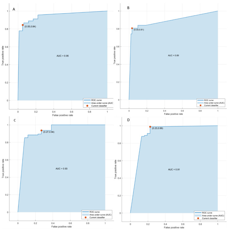
The aim of this study was to assess the efficacy of radiomics features obtained by EOB-MRI phase in order to predict clinical outcomes following liver resection in Colorectal Liver Metastases Patients, and evaluate recurrence, mutational status, pathological characteristic (mucinous) and surgical resection margin. This retrospective analysis was approved by the local Ethical Committee board of National Cancer of Naples, IRCCS "Fondazione Pascale". Radiological databases were interrogated from January 2018 to May 2021 in order to select patients with liver metastases with pathological proof and EOB-MRI study in pre-surgical setting. The cohort of patients included a training set (51 patients with 61 years of median age and 121 liver metastases) and an external validation set (30 patients with single lesion with 60 years of median age). For each segmented volume of interest by 2 expert radiologists, 851 radiomics features were extracted as median values using PyRadiomics. non-parametric test, intraclass correlation, receiver operating characteristic (ROC) analysis, linear regression modelling and pattern recognition methods (support vector machine (SVM), k-nearest neighbors (KNN), artificial neural network (NNET), and decision tree (DT)) were considered. The best predictor to discriminate expansive versus infiltrative front of tumor growth was HLH_glcm_MaximumProbability extraxted on VIBE_FA30 with an accuracy of 84%, a sensitivity of 83%, and a specificity of 82%. The best predictor to discriminate tumor budding was Inverse Variance obtained by the original GLCM matrix extraxted on VIBE_FA30 with an accuracy of 89%, a sensitivity of 96% and a specificity of 65%. The best predictor to differentiate the mucinous type of tumor was the HHL_glszm_ZoneVariance extraxted on VIBE_FA30 with an accuracy of 85%, a sensitivity of 46% and a specificity of 95%. The best predictor to identify tumor recurrence was the LHL_glcm_Correlation extraxted on VIBE_FA30 with an accuracy of 86%, a sensitivity of 52% and a specificity of 97%. The best linear regression model was obtained in the identification of the tumor growth front considering the height textural significant metrics by VIBE_FA10 (an accuracy of 89%; sensitivity of 93% and a specificity of 82%). Considering significant texture metrics tested with pattern recognition approaches, the best performance for each outcome was reached by a KNN in the identification of recurrence with the 3 textural significant features extracted by VIBE_FA10 (AUC of 91%, an accuracy of 93%; sensitivity of 99% and a specificity of 77%). Ours results confirmed the capacity of radiomics to identify as biomarkers, several prognostic features that could affect the treatment choice in patients with liver metastases, in order to obtain a more personalized approach.

摘要

本研究旨在评估EOB-MRI各期获得的影像组学特征预测结直肠癌肝转移患者肝切除术后临床结局的效能,并评估复发情况、突变状态、病理特征(黏液性)和手术切缘。这项回顾性分析得到了那不勒斯国家癌症研究所IRCCS“帕斯卡尔基金会”当地伦理委员会的批准。查询了2018年1月至2021年5月的放射学数据库,以选择有病理证据且术前进行过EOB-MRI检查的肝转移患者。患者队列包括一个训练集(51例患者,中位年龄61岁,121个肝转移灶)和一个外部验证集(30例单发病变患者,中位年龄60岁)。对于2名专业放射科医生分割的每个感兴趣体积,使用PyRadiomics提取851个影像组学特征作为中位数。考虑了非参数检验、组内相关性、受试者操作特征(ROC)分析、线性回归建模和模式识别方法(支持向量机(SVM)、k近邻(KNN)、人工神经网络(NNET)和决策树(DT))。区分肿瘤生长的膨胀性与浸润性前沿的最佳预测指标是在VIBE_FA30上提取的HLH_glcm_MaximumProbability,准确率为84%,灵敏度为83%,特异度为82%。区分肿瘤芽生的最佳预测指标是在VIBE_FA30上提取的原始GLCM矩阵的Inverse Variance,准确率为89%,灵敏度为96%,特异度为65%。区分黏液性肿瘤类型的最佳预测指标是在VIBE_FA30上提取的HHL_glszm_ZoneVariance,准确率为85%,灵敏度为46%,特异度为95%。识别肿瘤复发的最佳预测指标是在VIBE_FA30上提取的LHL_glcm_Correlation,准确率为86%,灵敏度为52%,特异度为97%。在考虑VIBE_FA10的高度纹理显著指标识别肿瘤生长前沿时获得了最佳线性回归模型(准确率89%;灵敏度93%,特异度82%)。考虑用模式识别方法测试的显著纹理指标,在识别复发时,KNN使用VIBE_FA10提取的3个纹理显著特征达到了每个结局的最佳性能(AUC为91%,准确率93%;灵敏度99%,特异度77%)。我们的结果证实了影像组学能够识别作为生物标志物的几种预后特征,这些特征可能会影响肝转移患者的治疗选择,从而获得更个性化的治疗方法。

https://cdn.ncbi.nlm.nih.gov/pmc/blobs/fa21/8909637/9dd82e3ad1de/cancers-14-01239-g006.jpg
https://cdn.ncbi.nlm.nih.gov/pmc/blobs/fa21/8909637/a30c226872a3/cancers-14-01239-g001.jpg
https://cdn.ncbi.nlm.nih.gov/pmc/blobs/fa21/8909637/2608274adc8f/cancers-14-01239-g002.jpg
https://cdn.ncbi.nlm.nih.gov/pmc/blobs/fa21/8909637/c87cb2011bbb/cancers-14-01239-g003.jpg
https://cdn.ncbi.nlm.nih.gov/pmc/blobs/fa21/8909637/2ebae274564b/cancers-14-01239-g004.jpg
https://cdn.ncbi.nlm.nih.gov/pmc/blobs/fa21/8909637/280b104d6247/cancers-14-01239-g005.jpg
https://cdn.ncbi.nlm.nih.gov/pmc/blobs/fa21/8909637/9dd82e3ad1de/cancers-14-01239-g006.jpg

相似文献

1
EOB-MR Based Radiomics Analysis to Assess Clinical Outcomes following Liver Resection in Colorectal Liver Metastases.基于增强外泌体磁共振成像的放射组学分析评估结直肠癌肝转移肝切除术后的临床结局
Cancers (Basel). 2022 Feb 27;14(5):1239. doi: 10.3390/cancers14051239.
2
Contrast MR-Based Radiomics and Machine Learning Analysis to Assess Clinical Outcomes following Liver Resection in Colorectal Liver Metastases: A Preliminary Study.基于对比增强磁共振成像的影像组学与机器学习分析评估结直肠癌肝转移肝切除术后的临床结局:一项初步研究
Cancers (Basel). 2022 Feb 22;14(5):1110. doi: 10.3390/cancers14051110.
3
Radiomics textural features by MR imaging to assess clinical outcomes following liver resection in colorectal liver metastases.通过磁共振成像的影像组学纹理特征评估结直肠癌肝转移肝切除术后的临床结局。
Radiol Med. 2022 May;127(5):461-470. doi: 10.1007/s11547-022-01477-6. Epub 2022 Mar 26.
4
CT-Based Radiomics Analysis to Predict Histopathological Outcomes Following Liver Resection in Colorectal Liver Metastases.基于CT的影像组学分析预测结直肠癌肝转移肝切除术后的组织病理学结果
Cancers (Basel). 2022 Mar 24;14(7):1648. doi: 10.3390/cancers14071648.
5
Machine Learning and Radiomics Analysis for Tumor Budding Prediction in Colorectal Liver Metastases Magnetic Resonance Imaging Assessment.用于结直肠癌肝转移磁共振成像评估中肿瘤芽生预测的机器学习与影像组学分析
Diagnostics (Basel). 2024 Jan 9;14(2):152. doi: 10.3390/diagnostics14020152.
6
Radiomics and machine learning analysis based on magnetic resonance imaging in the assessment of liver mucinous colorectal metastases.基于磁共振成像的放射组学和机器学习分析在评估肝黏液性结直肠转移中的应用。
Radiol Med. 2022 Jul;127(7):763-772. doi: 10.1007/s11547-022-01501-9. Epub 2022 Jun 2.
7
Radiomics and machine learning analysis by computed tomography and magnetic resonance imaging in colorectal liver metastases prognostic assessment.基于计算机断层扫描和磁共振成像的影像组学与机器学习分析在结直肠癌肝转移预后评估中的应用
Radiol Med. 2023 Nov;128(11):1310-1332. doi: 10.1007/s11547-023-01710-w. Epub 2023 Sep 11.
8
Radiomics and Machine Learning Analysis Based on Magnetic Resonance Imaging in the Assessment of Colorectal Liver Metastases Growth Pattern.基于磁共振成像的影像组学和机器学习分析在评估结直肠癌肝转移生长模式中的应用
Diagnostics (Basel). 2022 Apr 29;12(5):1115. doi: 10.3390/diagnostics12051115.
9
Machine learning-based radiomics analysis in predicting RAS mutational status using magnetic resonance imaging.基于机器学习的放射组学分析利用磁共振成像预测 RAS 突变状态。
Radiol Med. 2024 Mar;129(3):420-428. doi: 10.1007/s11547-024-01779-x. Epub 2024 Feb 2.
10
Radiomics and Artificial Intelligence Analysis with Textural Metrics Extracted by Contrast-Enhanced Mammography in the Breast Lesions Classification.通过对比增强乳腺钼靶摄影提取纹理特征的放射组学和人工智能分析在乳腺病变分类中的应用
Diagnostics (Basel). 2021 Apr 30;11(5):815. doi: 10.3390/diagnostics11050815.

引用本文的文献

1
Radiomics in radiology: What the radiologist needs to know about technical aspects and clinical impact.放射学中的放射组学:放射科医生需要了解的技术方面和临床影响。
Radiol Med. 2024 Dec;129(12):1751-1765. doi: 10.1007/s11547-024-01904-w. Epub 2024 Oct 30.
2
Artificial intelligence in predicting recurrence after first-line treatment of liver cancer: a systematic review and meta-analysis.人工智能在预测肝癌一线治疗后复发中的应用:系统评价和荟萃分析。
BMC Med Imaging. 2024 Oct 7;24(1):263. doi: 10.1186/s12880-024-01440-z.
3
Machine learning and radiomics analysis by computed tomography in colorectal liver metastases patients for RAS mutational status prediction.

本文引用的文献

1
CT radiomics models are unable to predict new liver metastasis after successful thermal ablation of colorectal liver metastases.CT 放射组学模型无法预测结直肠癌肝转移热消融治疗后新发肝转移。
Acta Radiol. 2023 Jan;64(1):5-12. doi: 10.1177/02841851211060437. Epub 2021 Dec 17.
2
Delta radiomics: a systematic review.德尔塔放射组学:系统评价。
Radiol Med. 2021 Dec;126(12):1571-1583. doi: 10.1007/s11547-021-01436-7. Epub 2021 Dec 4.
3
Automatic PI-RADS assignment by means of formal methods.通过形式化方法进行自动 PI-RADS 赋值。
基于 CT 的机器学习和放射组学分析预测结直肠癌肝转移患者的 RAS 基因突变状态。
Radiol Med. 2024 Jul;129(7):957-966. doi: 10.1007/s11547-024-01828-5. Epub 2024 May 18.
4
Preliminary data on artificial intelligence tool in magnetic resonance imaging assessment of degenerative pathologies of lumbar spine.人工智能工具在腰椎退行性病变磁共振成像评估中的初步数据。
Radiol Med. 2024 Apr;129(4):623-630. doi: 10.1007/s11547-024-01791-1. Epub 2024 Feb 13.
5
Machine learning-based radiomics analysis in predicting RAS mutational status using magnetic resonance imaging.基于机器学习的放射组学分析利用磁共振成像预测 RAS 突变状态。
Radiol Med. 2024 Mar;129(3):420-428. doi: 10.1007/s11547-024-01779-x. Epub 2024 Feb 2.
6
Machine Learning and Radiomics Analysis for Tumor Budding Prediction in Colorectal Liver Metastases Magnetic Resonance Imaging Assessment.用于结直肠癌肝转移磁共振成像评估中肿瘤芽生预测的机器学习与影像组学分析
Diagnostics (Basel). 2024 Jan 9;14(2):152. doi: 10.3390/diagnostics14020152.
7
Prognostic Assessment of Gastropancreatic Neuroendocrine Neoplasm: Prospects and Limits of Radiomics.胃胰腺神经内分泌肿瘤的预后评估:影像组学的前景与局限
Diagnostics (Basel). 2023 Sep 7;13(18):2877. doi: 10.3390/diagnostics13182877.
8
Multifaceted Insights into Innovative Approaches to Treating Colorectal Cancer Metastasis: From Emerging Biological Factors to Radiomics.结直肠癌转移治疗创新方法的多方面见解:从新兴生物因子到影像组学
Cancers (Basel). 2023 Sep 20;15(18):4644. doi: 10.3390/cancers15184644.
9
Radiomics and machine learning analysis by computed tomography and magnetic resonance imaging in colorectal liver metastases prognostic assessment.基于计算机断层扫描和磁共振成像的影像组学与机器学习分析在结直肠癌肝转移预后评估中的应用
Radiol Med. 2023 Nov;128(11):1310-1332. doi: 10.1007/s11547-023-01710-w. Epub 2023 Sep 11.
10
ctDNA as a prognostic biomarker in resectable CLM: Systematic review and meta-analysis.循环肿瘤DNA作为可切除性结直肠癌肝转移预后生物标志物的系统评价与Meta分析
Open Life Sci. 2023 May 19;18(1):20220615. doi: 10.1515/biol-2022-0615. eCollection 2023.
Radiol Med. 2022 Jan;127(1):83-89. doi: 10.1007/s11547-021-01431-y. Epub 2021 Nov 25.
4
A non-invasive, automated diagnosis of Menière's disease using radiomics and machine learning on conventional magnetic resonance imaging: A multicentric, case-controlled feasibility study.基于常规磁共振成像的放射组学和机器学习对梅尼埃病进行非侵入性、自动化诊断:一项多中心、病例对照可行性研究。
Radiol Med. 2022 Jan;127(1):72-82. doi: 10.1007/s11547-021-01425-w. Epub 2021 Nov 25.
5
Radiogenomics: Hunting Down Liver Metastasis in Colorectal Cancer Patients.放射基因组学:探寻结直肠癌患者的肝转移情况
Cancers (Basel). 2021 Nov 5;13(21):5547. doi: 10.3390/cancers13215547.
6
Preliminary Report on Computed Tomography Radiomics Features as Biomarkers to Immunotherapy Selection in Lung Adenocarcinoma Patients.计算机断层扫描影像组学特征作为肺腺癌患者免疫治疗选择生物标志物的初步报告
Cancers (Basel). 2021 Aug 7;13(16):3992. doi: 10.3390/cancers13163992.
7
A deep look into radiomics.深入探讨放射组学。
Radiol Med. 2021 Oct;126(10):1296-1311. doi: 10.1007/s11547-021-01389-x. Epub 2021 Jul 2.
8
Radiomics in hepatic metastasis by colorectal cancer.结直肠癌肝转移的影像组学
Infect Agent Cancer. 2021 Jun 2;16(1):39. doi: 10.1186/s13027-021-00379-y.
9
Radiomics and Artificial Intelligence Analysis with Textural Metrics Extracted by Contrast-Enhanced Mammography in the Breast Lesions Classification.通过对比增强乳腺钼靶摄影提取纹理特征的放射组学和人工智能分析在乳腺病变分类中的应用
Diagnostics (Basel). 2021 Apr 30;11(5):815. doi: 10.3390/diagnostics11050815.
10
Interventional Radiology ex-machina: impact of Artificial Intelligence on practice.介入放射学的救星:人工智能对实践的影响。
Radiol Med. 2021 Jul;126(7):998-1006. doi: 10.1007/s11547-021-01351-x. Epub 2021 Apr 16.