Molinero-Mourelle Pedro, Cascos-Sanchez Rocio, Yilmaz Burak, Lam Walter Yu Hang, Pow Edmond Ho Nang, Del Río Highsmith Jaime, Gómez-Polo Miguel
Department of Conservative Dentistry and Orofacial Prosthodontics, Faculty of Dentistry, Complutense University of Madrid, 28040 Madrid, Spain.
Department of Reconstructive Dentistry and Gerodontology, School of Dental Medicine, University of Bern, 3007 Bern, Switzerland.
Materials (Basel). 2021 Apr 30;14(9):2348. doi: 10.3390/ma14092348.
The aim of this in vitro study was to investigate the microgaps at the implant-abutment interface when zirconia (Zr) and CAD/CAM or cast Co-Cr abutments were used.
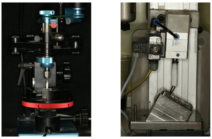
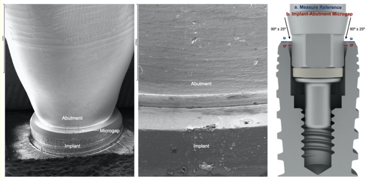
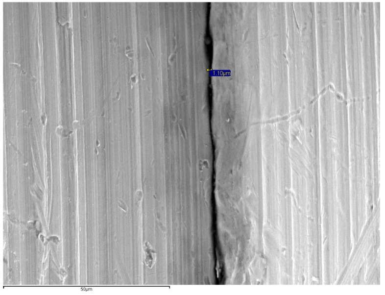
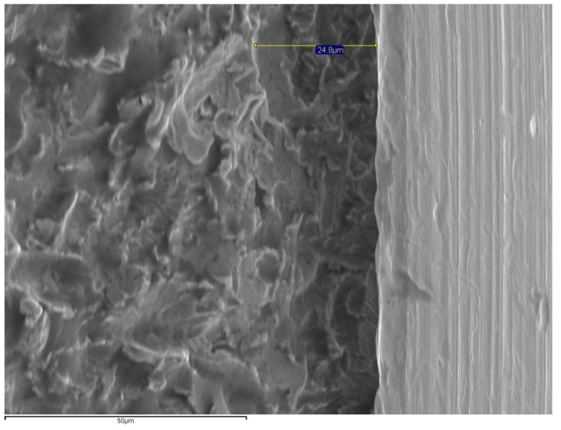
Sixty-four conical connection implants and their abutments were divided into four groups (Co-Cr (milled, laser-sintered and castable) and Zirconia (milled)). After chewing simulation (300,000 cycles, under 200 N loads at 2 Hz at a 30° angle) and thermocycling (10,000 cycles, 5 to 50 °C, dwelling time 55 s), the implant-abutment microgap was measured 14 times at each of the four anatomical aspects on each specimen by using a scanning electron microscope (SEM). Kruskal-Wallis and pair-wise comparison were used to analyze the data (α = 0.05).
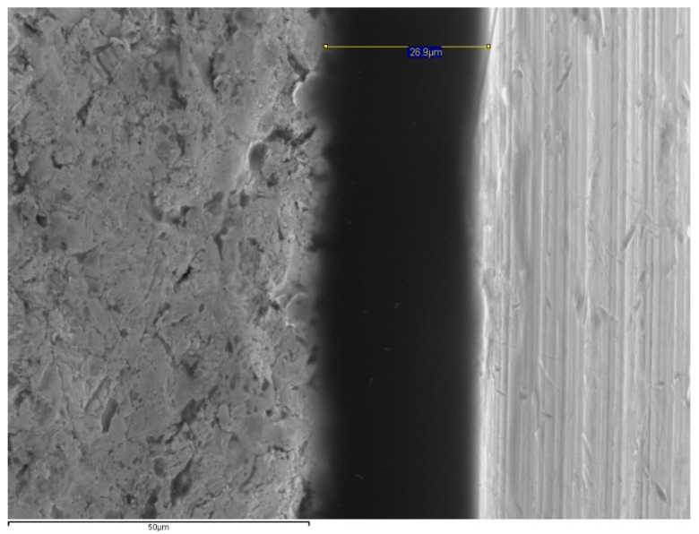
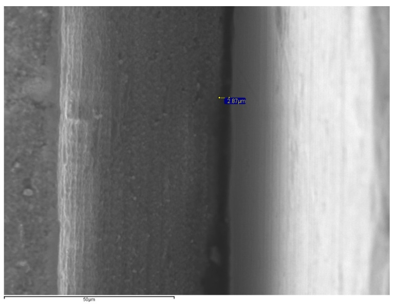
The SEM analysis revealed smaller microgaps with Co-Cr milled abutments (0.69-8.39 μm) followed by Zr abutments (0.12-6.57 μm), Co-Cr sintered (7.31-25.7 μm) and cast Co-Cr (1.68-85.97 μm). Statistically significant differences were found between milled and cast Co-Cr, milled and laser-sintered Co-Cr, and between Zr and cast and laser-sintered Co-Cr ( < 0.05).
The material and the abutment fabrication technique affected the implant-abutment microgap magnitude. The Zr and the milled Co-Cr presented smaller microgaps. Although the CAD/CAM abutments presented the most favorable values, all tested groups had microgaps within a range of 10 to 150 μm.
本体外研究的目的是调查使用氧化锆(Zr)以及计算机辅助设计/计算机辅助制造(CAD/CAM)或铸造钴铬基台时种植体-基台界面处的微间隙。
将64个锥形连接种植体及其基台分为四组(钴铬(铣削、激光烧结和可铸造)和氧化锆(铣削))。在咀嚼模拟(300,000次循环,在2赫兹频率、200牛负载、30°角条件下)和热循环(10,000次循环,5至50°C,停留时间55秒)后,使用扫描电子显微镜(SEM)在每个标本的四个解剖部位各测量14次种植体-基台微间隙。采用Kruskal-Wallis检验和两两比较分析数据(α = 0.05)。
SEM分析显示,钴铬铣削基台的微间隙较小(0.69 - 8.39微米),其次是氧化锆基台(0.12 - 6.57微米)、钴铬烧结基台(7.31 - 25.7微米)和铸造钴铬基台(1.68 - 85.97微米)。在铣削钴铬与铸造钴铬、铣削钴铬与激光烧结钴铬以及氧化锆与铸造和激光烧结钴铬之间发现有统计学显著差异(P < 0.05)。
材料和基台制作技术影响种植体-基台微间隙大小。氧化锆和铣削钴铬的微间隙较小。尽管CAD/CAM基台呈现出最有利的值,但所有测试组的微间隙都在10至150微米范围内。