• 文献检索
  • 文档翻译
  • 深度研究
  • 学术资讯
  • Suppr Zotero 插件Zotero 插件
  • 邀请有礼
  • 套餐&价格
  • 历史记录
应用&插件
Suppr Zotero 插件Zotero 插件浏览器插件Mac 客户端Windows 客户端微信小程序
定价
高级版会员购买积分包购买API积分包
服务
文献检索文档翻译深度研究API 文档MCP 服务
关于我们
关于 Suppr公司介绍联系我们用户协议隐私条款
关注我们

Suppr 超能文献

核心技术专利:CN118964589B侵权必究
粤ICP备2023148730 号-1Suppr @ 2026

文献检索

告别复杂PubMed语法,用中文像聊天一样搜索,搜遍4000万医学文献。AI智能推荐,让科研检索更轻松。

立即免费搜索

文件翻译

保留排版,准确专业,支持PDF/Word/PPT等文件格式,支持 12+语言互译。

免费翻译文档

深度研究

AI帮你快速写综述,25分钟生成高质量综述,智能提取关键信息,辅助科研写作。

立即免费体验

简化立体定向放射外科治疗的颅内病变线性尺寸的肿瘤体积估计

Simplifying Tumor Volume Estimation from Linear Dimensions for Intra-Cranial Lesions Treated with Stereotactic Radiosurgery.

作者信息

Singhal Sakshi, Gill Maneet, Srivastava Chinmaya, Gupta Darpan, Kumar Ashok, Kaushik Aruna, Semwal Manoj Kumar

机构信息

Division of PET Imaging, Institute of Nuclear Medicine and Allied Sciences, Delhi, India.

Department of Neurosurgery, Army Hospital Research and Referral, Delhi, India.

出版信息

J Med Phys. 2020 Oct-Dec;45(4):199-205. doi: 10.4103/jmp.JMP_56_20. Epub 2021 Feb 2.

DOI:10.4103/jmp.JMP_56_20
PMID:33953494
原文链接:https://pmc.ncbi.nlm.nih.gov/articles/PMC8074724/
Abstract

AIMS

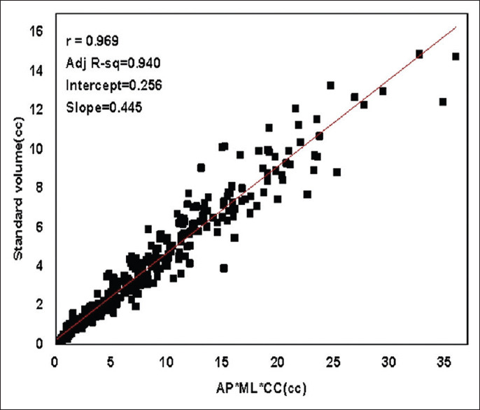
This study aims to derive simple yet robust formula(s) for the calculation of cranial tumor volume using linear tumor dimensions in anterioposterior (AP), mediolateral (ML) and craniocaudal (CC) directions and also propose a reproducible methodology for tumor dimension measurements.

MATERIALS AND METHODS

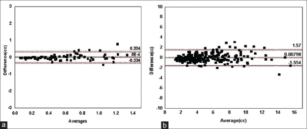
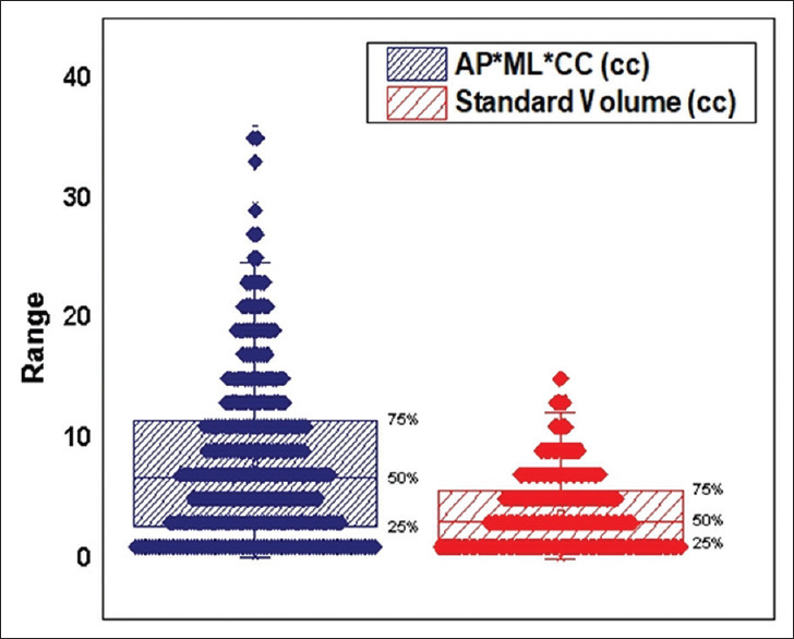
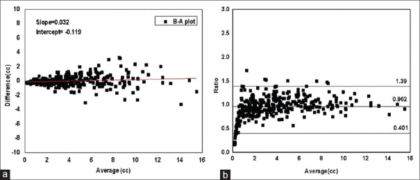
Magnetic resonance images (MRI) of 337 patients planned for Gammaknife Stereotactic Radiosurgery for different types of brain tumors were analyzed using Leksell Gamma Plan (LGP) software. Tumor volume in three dimensional was outlined and maximum tumor diameters were measured in three orthogonal directions AP, ML, and CC on the MRI. Formulas were derived to calculate tumor volume from AP, ML, and CC diameters using linear regression technique. An agreement between the calculated volume and standard volume observed from LGP software was determined using Bland Altman (B-A) plot. A comparison was made between the volume calculated using traditionally used formula of ellipsoid, standard volume obtained from LGP software and volume calculated from formulas derived in the present study.

RESULTS

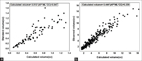
The tumors were divided into two categories based on their size for better volume prediction. The tumors having product of their diameters in the range 0-2.5cc were called "small tumors" and the formula proposed for their volume estimation ( + 0.047 ) was found to predict the tumor volume with an average bias of 0.0005cc. For "large tumors," having product of diameters in the range 2.5-36cc, the proposed formula ( + 0.339 ) predicted the tumor volume with an average bias of 0.007cc.

CONCLUSIONS

The two formulas proposed in the study are more accurate as compared to the commonly used formula that considers the tumors as ellipsoids. The methodology proposed in the study for measurement of linear tumor dimensions is simple and reproducible.

摘要

目的

本研究旨在推导简单而可靠的公式,用于根据前后径(AP)、内外径(ML)和颅尾径(CC)方向的线性肿瘤尺寸计算颅部肿瘤体积,并提出一种可重复的肿瘤尺寸测量方法。

材料与方法

使用Leksell伽马计划(LGP)软件分析了337例计划接受伽马刀立体定向放射外科治疗不同类型脑肿瘤患者的磁共振成像(MRI)。在MRI上勾勒出三维肿瘤体积,并在三个正交方向AP、ML和CC上测量最大肿瘤直径。使用线性回归技术推导从AP、ML和CC直径计算肿瘤体积的公式。使用布兰德-奥特曼(B-A)图确定计算体积与LGP软件观察到的标准体积之间的一致性。对使用传统椭球体公式计算的体积、从LGP软件获得的标准体积以及本研究推导公式计算的体积进行比较。

结果

根据肿瘤大小将其分为两类以更好地预测体积。直径乘积在0 - 2.5立方厘米范围内的肿瘤称为“小肿瘤”,发现为其体积估计提出的公式(+ 0.047)预测肿瘤体积的平均偏差为0.0005立方厘米。对于直径乘积在2.5 - 36立方厘米范围内的“大肿瘤”,提出的公式(+ 0.339)预测肿瘤体积的平均偏差为0.007立方厘米。

结论

与将肿瘤视为椭球体的常用公式相比,本研究提出的两个公式更准确。本研究提出的测量线性肿瘤尺寸的方法简单且可重复。

https://cdn.ncbi.nlm.nih.gov/pmc/blobs/5568/8074724/bfb170f3a8e7/JMP-45-199-g007.jpg
https://cdn.ncbi.nlm.nih.gov/pmc/blobs/5568/8074724/51d010b29ce9/JMP-45-199-g003.jpg
https://cdn.ncbi.nlm.nih.gov/pmc/blobs/5568/8074724/ff0058aa48f1/JMP-45-199-g004.jpg
https://cdn.ncbi.nlm.nih.gov/pmc/blobs/5568/8074724/291d40eb7e60/JMP-45-199-g005.jpg
https://cdn.ncbi.nlm.nih.gov/pmc/blobs/5568/8074724/2044e5c9f11f/JMP-45-199-g006.jpg
https://cdn.ncbi.nlm.nih.gov/pmc/blobs/5568/8074724/bfb170f3a8e7/JMP-45-199-g007.jpg

相似文献

1
Simplifying Tumor Volume Estimation from Linear Dimensions for Intra-Cranial Lesions Treated with Stereotactic Radiosurgery.简化立体定向放射外科治疗的颅内病变线性尺寸的肿瘤体积估计
J Med Phys. 2020 Oct-Dec;45(4):199-205. doi: 10.4103/jmp.JMP_56_20. Epub 2021 Feb 2.
2
Prediction of volumetric data errors in patients treated with gamma knife radiosurgery.伽玛刀放射外科治疗患者容积数据误差的预测。
Stereotact Funct Neurosurg. 2007;85(4):184-91. doi: 10.1159/000101297. Epub 2007 Mar 27.
3
Assessment of lung cancer response after nonoperative therapy: tumor diameter, bidimensional product, and volume. A serial CT scan-based study.非手术治疗后肺癌反应的评估:肿瘤直径、二维乘积和体积。一项基于CT扫描序列的研究。
Int J Radiat Oncol Biol Phys. 2001 Sep 1;51(1):56-61. doi: 10.1016/s0360-3016(01)01615-7.
4
Adaptive hybrid surgery analysis (AHSA) for adjuvant gamma knife radiosurgery treatment of vestibular schwannoma residuals.用于前庭神经鞘瘤残余瘤辅助伽玛刀放射外科治疗的自适应混合手术分析(AHSA)
Clin Neurol Neurosurg. 2019 Oct;185:105487. doi: 10.1016/j.clineuro.2019.105487. Epub 2019 Aug 12.
5
Mammographic determination of breast volume by elliptical cone estimation.用椭圆锥估计法进行乳腺体积的 X 光摄影测定。
World J Surg. 2010 Jul;34(7):1442-5. doi: 10.1007/s00268-009-0283-0.
6
Clinical significance of 3D reconstruction of arteriovenous malformation using digital subtraction angiography and its modification with CT information in stereotactic radiosurgery.数字减影血管造影对动静脉畸形进行三维重建及其在立体定向放射外科中结合CT信息进行修正的临床意义
Int J Radiat Oncol Biol Phys. 2003 Dec 1;57(5):1392-9. doi: 10.1016/s0360-3016(03)00780-6.
7
Splenic volume model constructed from standardized one-dimensional MDCT measurements.基于标准化一维 MDCT 测量构建的脾脏体积模型。
AJR Am J Roentgenol. 2011 Feb;196(2):367-72. doi: 10.2214/AJR.10.4453.
8
The Application of Magnetic Resonance Imaging-Deformed 11C-Methionine-Positron Emission Tomography Images in Stereotactic Radiosurgery.磁共振成像-变形11C-蛋氨酸-正电子发射断层扫描图像在立体定向放射外科中的应用
Stereotact Funct Neurosurg. 2019;97(4):217-224. doi: 10.1159/000503732. Epub 2019 Nov 6.
9
Magnetic resonance imaging characteristics and the prediction of outcome of vestibular schwannomas following Gamma Knife radiosurgery.伽玛刀放射外科治疗前庭神经鞘瘤后的磁共振成像特征及其对预后的预测。
J Neurosurg. 2017 Dec;127(6):1384-1391. doi: 10.3171/2016.9.JNS161510. Epub 2017 Feb 10.
10
Verification of dose profiles generated by the convolution algorithm of the gamma knife radiosurgery planning system.伽玛刀放射外科计划系统卷积算法生成剂量分布的验证。
Med Phys. 2017 Sep;44(9):4880-4889. doi: 10.1002/mp.12347. Epub 2017 Jul 28.

引用本文的文献

1
Epilepsy in Association with Giant Skull Base Tumors: A Retrospective Case Series of Giant Skull Base Tumors in the Anterior and Middle Fossa.与巨大颅底肿瘤相关的癫痫:前颅窝和中颅窝巨大颅底肿瘤的回顾性病例系列
J Neurol Surg B Skull Base. 2024 May 3;86(3):254-261. doi: 10.1055/a-2297-8981. eCollection 2025 Jun.
2
Excellent Response to Trastuzumab Deruxtecan of a Large Medullary Metastasis from Human Epidermal Growth Factor Receptor 2-Positive Breast Cancer: A Case Report.曲妥珠单抗德鲁替康对人表皮生长因子受体2阳性乳腺癌大髓样转移灶的卓越疗效:一例报告
Case Rep Oncol. 2025 Mar 29;18(1):523-530. doi: 10.1159/000545577. eCollection 2025 Jan-Dec.
3

本文引用的文献

1
A Cancer Care Ontario Organizational Guideline for the Delivery of Stereotactic Radiosurgery for Brain Metastasis in Ontario, Canada.加拿大安大略省癌症护理组织的脑转移瘤立体定向放射外科治疗指南
Pract Radiat Oncol. 2020 Jul-Aug;10(4):243-254. doi: 10.1016/j.prro.2019.11.002. Epub 2019 Nov 26.
2
The growing importance of lesion volume as a prognostic factor in patients with multiple brain metastases treated with stereotactic radiosurgery.立体定向放射外科治疗多发性脑转移瘤患者中病灶体积作为预后因素的重要性日益增加。
Cancer Med. 2018 Mar;7(3):757-764. doi: 10.1002/cam4.1352. Epub 2018 Feb 14.
3
Calculating the Tumor Volumes in Vestibular Schwannomas: Are the ABC/2 and Volumetric Methods Comparable?
Chondromyxoid fibroma: A retrospective evaluation of 31 cases.
软骨黏液样纤维瘤:31 例回顾性评估。
Jt Dis Relat Surg. 2024 Apr 26;35(2):377-385. doi: 10.52312/jdrs.2024.1620.
4
Investigation of the effects of Gamma Knife radiosurgery on optic pathways using diffusion tensor MRI within the first year after treatment.探讨伽玛刀放射外科治疗后 1 年内应用弥散张量 MRI 对视路的影响。
Neuroradiology. 2024 Apr;66(4):609-620. doi: 10.1007/s00234-024-03296-0. Epub 2024 Feb 16.
5
Lung Micrometastases Display ECM Depletion and Softening While Macrometastases Are 30-Fold Stiffer and Enriched in Fibronectin.肺微转移灶表现出细胞外基质耗竭和软化,而大转移灶的硬度则是微转移灶的30倍,且富含纤连蛋白。
Cancers (Basel). 2023 Apr 21;15(8):2404. doi: 10.3390/cancers15082404.
计算前庭神经鞘瘤的肿瘤体积:ABC/2法和容积法是否具有可比性?
Otol Neurotol. 2017 Jul;38(6):889-894. doi: 10.1097/MAO.0000000000001423.
4
The radiosurgery fractionation quandary: single fraction or hypofractionation?放射外科的分次治疗难题:单次分割还是低分割?
Neuro Oncol. 2017 Apr 1;19(suppl_2):ii38-ii49. doi: 10.1093/neuonc/now301.
5
Reporting of method comparison studies: a review of advice, an assessment of current practice, and specific suggestions for future reports.方法比较研究报告:建议综述、当前实践评估及未来报告具体建议。
Br J Anaesth. 2016 Nov;117(5):569-575. doi: 10.1093/bja/aew320.
6
Reliability of the Size Evaluation Method for Meningiomas: Maximum Diameter, ABC/2 Formula, and Planimetry Method.脑膜瘤大小评估方法的可靠性:最大直径、ABC/2公式和平面积测量法
World Neurosurg. 2016 Oct;94:80-88. doi: 10.1016/j.wneu.2016.06.108. Epub 2016 Jul 2.
7
Measuring glioma volumes: A comparison of linear measurement based formulae with the manual image segmentation technique.测量胶质瘤体积:基于线性测量的公式与手动图像分割技术的比较。
J Cancer Res Ther. 2016 Jan-Mar;12(1):161-8. doi: 10.4103/0973-1482.153999.
8
Assessing size of pituitary adenomas: a comparison of qualitative and quantitative methods on MR.评估垂体腺瘤的大小:磁共振成像上定性和定量方法的比较
Acta Neurochir (Wien). 2016 Apr;158(4):677-683. doi: 10.1007/s00701-015-2699-7. Epub 2016 Jan 29.
9
Understanding Bland Altman analysis.理解布兰德-奥特曼分析。
Biochem Med (Zagreb). 2015 Jun 5;25(2):141-51. doi: 10.11613/BM.2015.015. eCollection 2015.
10
Preoperative tumor size at MRI predicts deep myometrial invasion, lymph node metastases, and patient outcome in endometrial carcinomas.磁共振成像(MRI)术前肿瘤大小可预测子宫内膜癌的肌层深部浸润、淋巴结转移及患者预后。
Int J Gynecol Cancer. 2015 Mar;25(3):459-66. doi: 10.1097/IGC.0000000000000367.