Neuroradiology Division, Department of Radiology, Newark Beth Israel Medical Center, Newark, NJ, USA.
Department of Neurosurgery, Ayer Neuroscience Institute, The Hospital of Central Connecticut, New Britain, CT, USA.
Br J Cancer. 2021 Aug;125(5):641-657. doi: 10.1038/s41416-021-01387-w. Epub 2021 May 6.
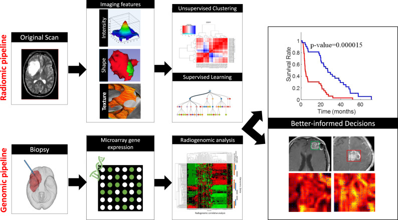
The natural history and treatment landscape of primary brain tumours are complicated by the varied tumour behaviour of primary or secondary gliomas (high-grade transformation of low-grade lesions), as well as the dilemmas with identification of radiation necrosis, tumour progression, and pseudoprogression on MRI. Radiomics and radiogenomics promise to offer precise diagnosis, predict prognosis, and assess tumour response to modern chemotherapy/immunotherapy and radiation therapy. This is achieved by a triumvirate of morphological, textural, and functional signatures, derived from a high-throughput extraction of quantitative voxel-level MR image metrics. However, the lack of standardisation of acquisition parameters and inconsistent methodology between working groups have made validations unreliable, hence multi-centre studies involving heterogenous study populations are warranted. We elucidate novel radiomic and radiogenomic workflow concepts and state-of-the-art descriptors in sub-visual MR image processing, with relevant literature on applications of such machine learning techniques in glioma management.
原发性脑肿瘤的自然史和治疗现状因原发性或继发性神经胶质瘤(低级病变的高级别转化)的不同肿瘤行为而变得复杂,此外,MRI 上还存在放射性坏死、肿瘤进展和假性进展的鉴别难题。放射组学和放射基因组学有望提供精确的诊断、预测预后,并评估肿瘤对现代化疗/免疫治疗和放疗的反应。这是通过从高通量提取定量体素级 MR 图像指标中得出的形态学、纹理和功能特征的三位一体来实现的。然而,由于采集参数缺乏标准化和工作组之间方法学的不一致,验证结果不可靠,因此需要涉及异质研究人群的多中心研究。我们阐述了亚视觉磁共振图像处理中新的放射组学和放射基因组学工作流程概念和最新描述符,并介绍了此类机器学习技术在神经胶质瘤管理中的应用相关文献。