Center for Global Infectious Disease Research, Seattle Children's Research Institute, Seattle, Washington, United States of America.
University of Washington Department of Microbiology, Seattle, Washington, United States of America.
PLoS Pathog. 2021 May 7;17(5):e1009575. doi: 10.1371/journal.ppat.1009575. eCollection 2021 May.
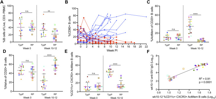
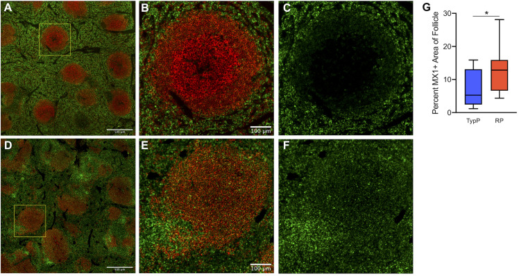
HIV-infected infants are at an increased risk of progressing rapidly to AIDS in the first weeks of life. Here, we evaluated immunological and virological parameters in 25 SIV-infected infant rhesus macaques to understand the factors influencing a rapid disease outcome. Infant macaques were infected with SIVmac251 and monitored for 10 to 17 weeks post-infection. SIV-infected infants were divided into either typical (TypP) or rapid (RP) progressor groups based on levels of plasma anti-SIV antibody and viral load, with RP infants having low SIV-specific antibodies and high viral loads. Following SIV infection, 11 out of 25 infant macaques exhibited an RP phenotype. Interestingly, TypP had lower levels of total CD4 T cells, similar reductions in CD4/CD8 ratios and elevated activation of CD8 T cells, as measured by the levels of HLA-DR, compared to RP. Differences between the two groups were identified in other immune cell populations, including a failure to expand activated memory (CD21-CD27+) B cells in peripheral blood in RP infant macaques, as well as reduced levels of germinal center (GC) B cells and T follicular helper (Tfh) cells in spleens (4- and 10-weeks post-SIV). Reduced B cell proliferation in splenic germinal GCs was associated with increased SIV+ cell density and follicular type 1 interferon (IFN)-induced immune activation. Further analyses determined that at 2-weeks post SIV infection TypP infants exhibited elevated levels of the GC-inducing chemokine CXCL13 in plasma, as well as significantly lower levels of viral envelope diversity compared to RP infants. Our findings provide evidence that early viral and immunologic events following SIV infection contributes to impairment of B cells, Tfh cells and germinal center formation, ultimately impeding the development of SIV-specific antibody responses in rapidly progressing infant macaques.
HIV 感染的婴儿在生命的最初几周内迅速发展为艾滋病的风险增加。在这里,我们评估了 25 只 SIV 感染的婴儿恒河猴的免疫和病毒学参数,以了解影响快速疾病结局的因素。婴儿恒河猴感染 SIVmac251 并在感染后 10 至 17 周进行监测。根据血浆抗 SIV 抗体和病毒载量,将 SIV 感染的婴儿分为典型(TypP)或快速(RP)进展组,RP 婴儿的 SIV 特异性抗体水平低,病毒载量高。SIV 感染后,25 只婴儿恒河猴中有 11 只表现出 RP 表型。有趣的是,与 RP 相比,TypP 的总 CD4 T 细胞水平较低,CD4/CD8 比值相似降低,CD8 T 细胞激活水平升高,如 HLA-DR 水平所示。两组之间在其他免疫细胞群体中存在差异,包括 RP 婴儿恒河猴外周血中激活的记忆性(CD21-CD27+)B 细胞无法扩增,以及脾脏中生发中心(GC)B 细胞和滤泡辅助性(Tfh)细胞水平降低(感染 SIV 后 4 周和 10 周)。脾脏生发中心 GC 中 B 细胞增殖减少与 SIV+细胞密度增加和滤泡型 1 型干扰素(IFN)诱导的免疫激活有关。进一步分析确定,在 SIV 感染后 2 周,TypP 婴儿的血浆中 GC 诱导趋化因子 CXCL13 水平升高,而与 RP 婴儿相比,病毒包膜多样性水平显著降低。我们的研究结果表明,SIV 感染后早期的病毒和免疫事件导致 B 细胞、Tfh 细胞和生发中心形成受损,最终阻碍了快速进展的婴儿恒河猴中 SIV 特异性抗体反应的发展。