Kark Sarah M, Birnie Matthew T, Baram Tallie Z, Yassa Michael A
Center for the Neurobiology of Learning and Memory, University of California, Irvine, Irvine, CA, United States.
Department of Neurobiology and Behavior, University of California, Irvine, Irvine, CA, United States.
Front Integr Neurosci. 2021 Apr 21;15:662293. doi: 10.3389/fnint.2021.662293. eCollection 2021.
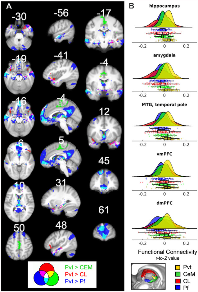
The paraventricular thalamic nucleus (PVT) is a small but highly connected nucleus of the dorsal midline thalamus. The PVT has garnered recent attention as a context-sensitive node within the thalamocortical arousal system that modulates state-dependent motivated behaviors. Once considered related to generalized arousal responses with non-specific impacts on behavior, accumulating evidence bolsters the contemporary view that discrete midline thalamic subnuclei belong to specialized corticolimbic and corticostriatal circuits related to attention, emotions, and cognition. However, the functional connectivity patterns of the human PVT have yet to be mapped. Here, we combined high-quality, high-resolution 7T and 3T resting state MRI data from 121 young adult participants from the Human Connectome Project (HCP) and thalamic subnuclei atlas masks to investigate resting state functional connectivity of the human PVT. The 7T results demonstrated extensive positive functional connectivity with the brainstem, midbrain, ventral and dorsal medial prefrontal cortex (mPFC), anterior and posterior cingulate, ventral striatum, hippocampus, and amygdala. These connections persist upon controlling for functional connectivity of the rest of the thalamus. Whole-brain contrasts provided further evidence that, compared to three nearby midline thalamic subnuclei, functional connectivity of the PVT is strong with the hippocampus, amygdala, ventral and dorsal mPFC, and middle temporal gyrus. These findings suggest that, even during rest, the human PVT is functionally coupled with many regions known to be structurally connected to rodent and non-human primate PVT. Further, cosine similarity analysis results suggested the PVT is integrated into the default mode network (DMN), an intrinsic connectivity network associated with episodic memory and self-referential thought. The current work provides a much-needed foundation for ongoing and future work examining the functional roles of the PVT in humans.
室旁丘脑核(PVT)是背侧中线丘脑的一个小但高度连接的核团。作为丘脑皮质觉醒系统中一个对环境敏感的节点,PVT最近受到了关注,该系统调节与状态相关的动机行为。PVT曾被认为与对行为有非特异性影响的全身性觉醒反应有关,越来越多的证据支持当代观点,即离散的中线丘脑亚核属于与注意力、情绪和认知相关的特殊皮质边缘和皮质纹状体回路。然而,人类PVT的功能连接模式尚未被绘制出来。在这里,我们结合了来自人类连接组计划(HCP)的121名年轻成年参与者的高质量、高分辨率7T和3T静息态MRI数据以及丘脑亚核图谱掩码,以研究人类PVT的静息态功能连接。7T结果显示,PVT与脑干、中脑、腹侧和背侧内侧前额叶皮质(mPFC)、前扣带回和后扣带回、腹侧纹状体、海马体和杏仁核有广泛的正功能连接。在控制丘脑其他部分的功能连接后,这些连接仍然存在。全脑对比进一步证明,与附近三个中线丘脑亚核相比,PVT与海马体、杏仁核、腹侧和背侧mPFC以及颞中回的功能连接很强。这些发现表明,即使在休息时,人类PVT在功能上也与许多已知与啮齿动物和非人类灵长类动物PVT有结构连接的区域耦合。此外,余弦相似性分析结果表明,PVT被整合到默认模式网络(DMN)中,这是一个与情景记忆和自我参照思维相关的内在连接网络。目前的工作为正在进行的和未来研究PVT在人类中的功能作用的工作提供了急需的基础。