Key Laboratory of Imaging Diagnosis and Minimally Invasive Interventional Research of Zhejiang Province, Lishui Hospital, Zhejiang University School of Medicine, Lishui, 323000, Zhejiang, China.
Institute of Pharmaceutics, College of Pharmaceutical Sciences, Zhejiang University, Hangzhou, 310058, China.
J Nanobiotechnology. 2021 May 10;19(1):132. doi: 10.1186/s12951-021-00870-z.
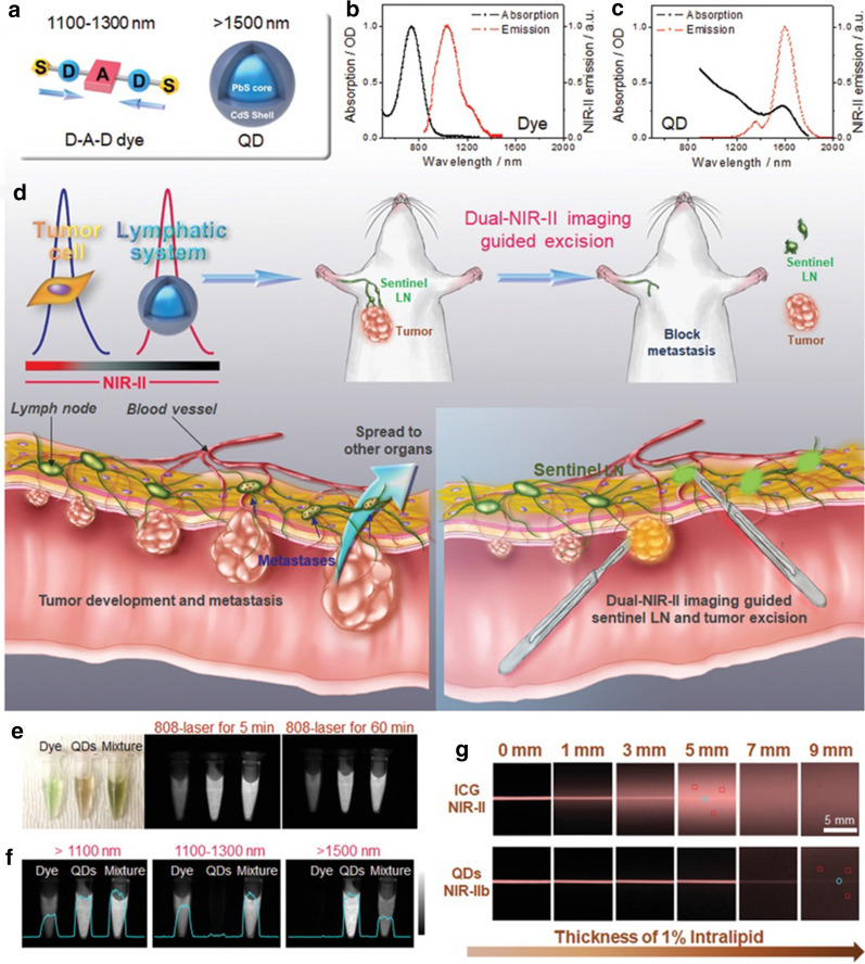
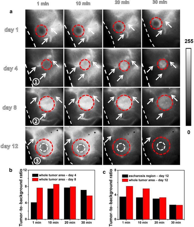
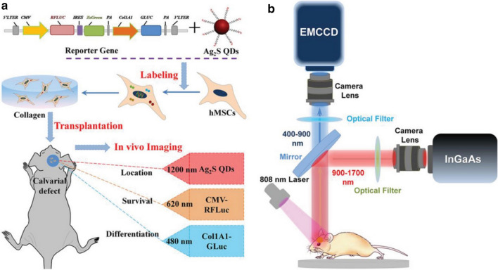
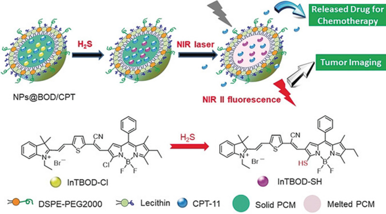
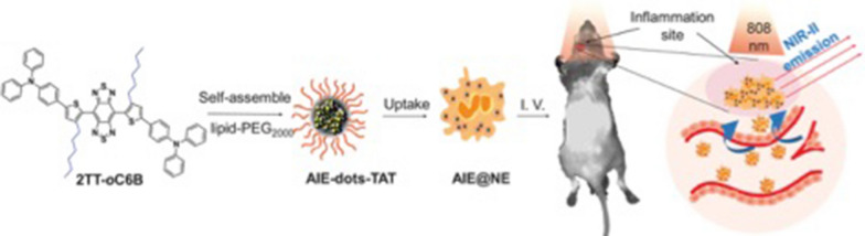
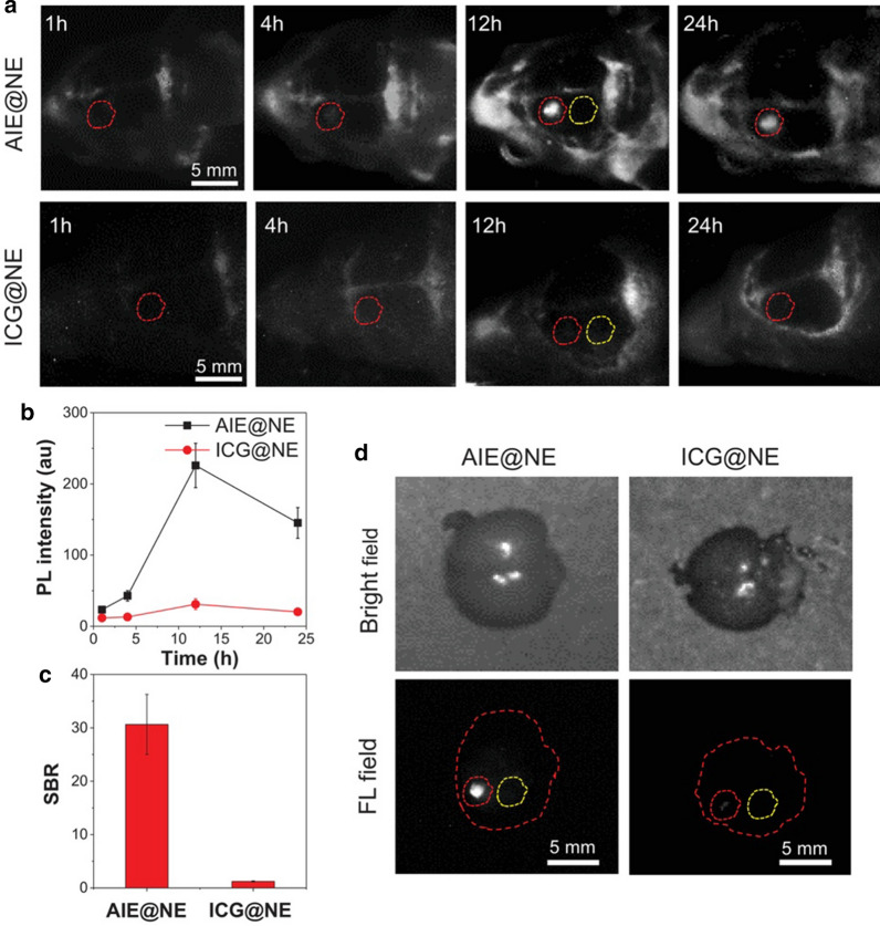
Molecular imaging technology enables us to observe the physiological or pathological processes in living tissue at the molecular level to accurately diagnose diseases at an early stage. Optical imaging can be employed to achieve the dynamic monitoring of tissue and pathological processes and has promising applications in biomedicine. The traditional first near-infrared (NIR-I) window (NIR-I, range from 700 to 900 nm) imaging technique has been available for more than two decades and has been extensively utilized in clinical diagnosis, treatment and scientific research. Compared with NIR-I, the second NIR window optical imaging (NIR-II, range from 1000 to 1700 nm) technology has low autofluorescence, a high signal-to-noise ratio, a high tissue penetration depth and a large Stokes shift. Recently, this technology has attracted significant attention and has also become a heavily researched topic in biomedicine. In this study, the optical characteristics of different fluorescence nanoprobes and the latest reports regarding the application of NIR-II nanoprobes in different biological tissues will be described. Furthermore, the existing problems and future application perspectives of NIR-II optical imaging probes will also be discussed.
分子成像技术使我们能够在分子水平上观察活体组织的生理或病理过程,从而能够及早准确诊断疾病。光学成像是实现组织和病理过程动态监测的一种手段,在生物医学领域具有广阔的应用前景。传统的近红外一区(NIR-I,700-900nm)成像技术已经有二十多年的历史,在临床诊断、治疗和科学研究中得到了广泛的应用。与 NIR-I 相比,近红外二区(NIR-II,1000-1700nm)光学成像技术具有自发荧光低、信噪比高、组织穿透深度大、斯托克斯位移大等优点。近年来,该技术受到了广泛关注,也成为生物医学领域的研究热点。本研究介绍了不同荧光纳米探针的光学特性以及 NIR-II 纳米探针在不同生物组织中的最新应用研究进展,同时对 NIR-II 光学成像探针存在的问题及未来应用前景进行了讨论。