Einarsson Emma, Svensson Jonas, Folkesson Elin, Kestilä Iida, Tjörnstrand Jon, Peterson Pernilla, Finnilä Mikko A J, Hughes H Velocity, Turkiewicz Aleksandra, Saarakkala Simo, Englund Martin
Medical Radiation Physics, Department of Translational Medicine, Lund University, Malmö, Sweden.
Clinical Epidemiology Unit, Orthopedics, Department of Clinical Sciences Lund, Lund University, Lund, Sweden.
Osteoarthr Cartil Open. 2020 Apr 3;2(2). doi: 10.1016/j.ocarto.2020.100061. eCollection 2020 Jun.
Quantitative magnetic resonance imaging (MRI), e.g. relaxation parameter mapping, may be sensitive to structural and compositional tissue changes, and could potentially be used to non-invasively detect and monitor early meniscus degeneration related to knee osteoarthritis.
To investigate MR relaxation times as potential biomarkers for meniscus degeneration through comparisons with histopathology.
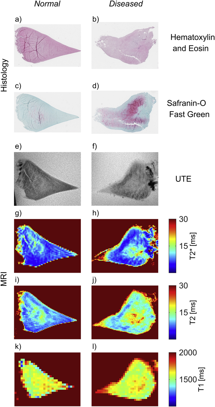
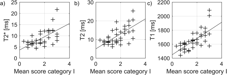
We measured MR relaxation parameters in the posterior horn of 40 menisci (medial and lateral) at a wide range of degenerative stages. T1, T2 and T2* were mapped using standard and ultrashort echo time sequences at 9.4 T and compared to gold standard histology using Pauli's histopathological scoring system, including assessment of surface integrity, collagen organization, cellularity and Safranin-O staining.
All three relaxation times increased with total Pauli score (mean difference per score (95% CI) for T2*: 0.62 (0.37, 0.86), T2: 0.83 (0.53, 1.1) and T1: 24.7 (16.5, 32.8) ms/score). Clear associations were seen with scores of surface integrity (mean difference per score for T2*: 3.0 (1.8, 4.2), T2: 4.0 (2.5, 5.5) and T1: 116 (75.6, 156) ms/score) and collagen organization (mean difference between highest and lowest score for T2*: 5.3 (1.6, 8.9), T2: 6.1 (1.7, 11) and T1: 204 (75.9, 332) ms). The results were less clear for the remaining histopathological measures.
MR relaxation times T1, T2 and T2* of human menisci are associated with histologically verified degenerative processes, in particular related to surface integrity and collagen organization. If confirmed MR relaxation times may thus be potential biomarkers for meniscus degeneration.
定量磁共振成像(MRI),例如弛豫参数映射,可能对组织的结构和成分变化敏感,并且有可能用于无创检测和监测与膝关节骨关节炎相关的早期半月板退变。
通过与组织病理学比较,研究磁共振弛豫时间作为半月板退变潜在生物标志物的情况。
我们在9.4T磁场下,使用标准和超短回波时间序列测量了40个半月板(内侧和外侧)后角在广泛退变阶段的磁共振弛豫参数。将T1、T2和T2*映射结果与使用保利组织病理学评分系统的金标准组织学结果进行比较,该评分系统包括对表面完整性、胶原组织、细胞密度和番红O染色的评估。
所有三个弛豫时间均随保利总分增加而增加(T2每分的平均差异(95%置信区间)为:0.62(0.37,0.86),T2为:0.83(0.53,1.1),T1为:24.7(16.5,32.8)ms/分)。表面完整性评分(T2每分的平均差异为:3.0(1.8,4.2),T2为:4.0(2.5,5.5),T1为:116(75.6,156)ms/分)和胶原组织评分(T2*最高与最低评分之间的平均差异为:5.3(1.6,8.9),T2为:6.1(1.7,11),T1为:204(75.9,332)ms)之间存在明显关联。对于其余组织病理学指标,结果不太明确。
人类半月板的磁共振弛豫时间T1、T2和T2*与经组织学证实的退变过程相关,尤其与表面完整性和胶原组织有关。如果得到证实,磁共振弛豫时间因此可能是半月板退变的潜在生物标志物。