College of Control Science and Engineering, China University of Petroleum (East China), Qingdao, China.
School of Computing, Newcastle University, Newcastle upon Tyne, UK.
Hum Brain Mapp. 2021 Aug 15;42(12):3777-3791. doi: 10.1002/hbm.25464. Epub 2021 May 11.
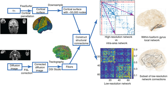
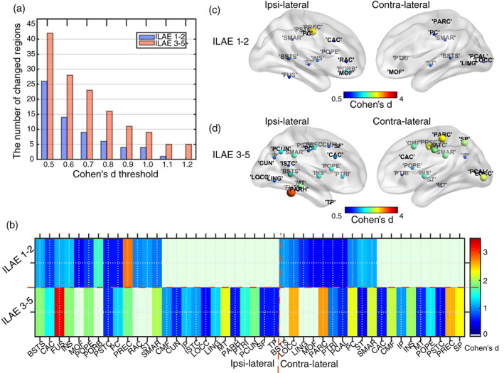
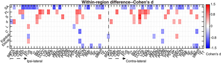
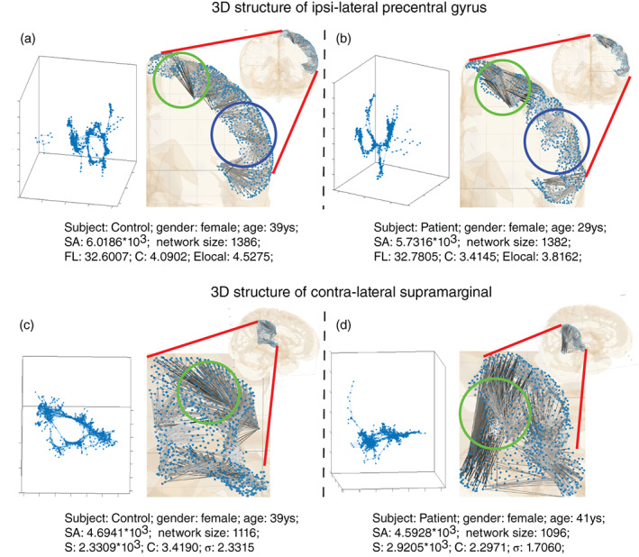
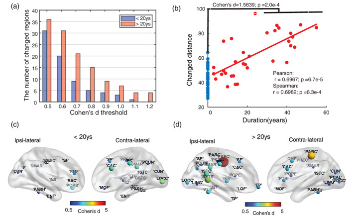
Finding clear connectome biomarkers for temporal lobe epilepsy (TLE) patients, in particular at early disease stages, remains a challenge. Currently, the whole-brain structural connectomes are analyzed based on coarse parcellations (up to 1,000 nodes). However, such global parcellation-based connectomes may be unsuitable for detecting more localized changes in patients. Here, we use a high-resolution network (50,000-nodes overall) to identify changes at the local level (within brain regions) and test its relation with duration and surgical outcome. Patients with TLE (n = 33) and age-, sex-matched healthy subjects (n = 36) underwent high-resolution (50,000 nodes) structural network construction based on deterministic tracking of diffusion tensor imaging. Nodes were allocated to 68 cortical regions according to the Desikan-Killany atlas. The connectivity within regions was then used to predict surgical outcome. MRI processing, network reconstruction, and visualization of network changes were integrated into the NICARA (https://nicara.eu). Lower clustering coefficient and higher edge density were found for local connectivity within regions in patients, but were absent for the global network between regions (68 cortical regions). Local connectivity changes, in terms of the number of changed regions and the magnitude of changes, increased with disease duration. Local connectivity yielded a better surgical outcome prediction (Mean value: 95.39% accuracy, 92.76% sensitivity, and 100% specificity) than global connectivity. Connectivity within regions, compared to structural connectivity between brain regions, can be a more efficient biomarker for epilepsy assessment and surgery outcome prediction of medically intractable TLE.
寻找明确的颞叶癫痫(TLE)患者连接组学生物标志物,尤其是在疾病早期阶段,仍然是一个挑战。目前,全脑结构连接组学是基于粗略分割(最多 1000 个节点)进行分析的。然而,这种基于全局分割的连接组可能不适合检测患者更局部的变化。在这里,我们使用高分辨率网络(总体约 50000 个节点)来识别局部水平(脑区内)的变化,并测试其与病程和手术结果的关系。33 名 TLE 患者(n=33)和年龄、性别匹配的健康对照者(n=36)接受了高分辨率(约 50000 个节点)结构网络构建,该网络基于扩散张量成像的确定性追踪。节点根据 Desikan-Killany 图谱分配到 68 个皮质区域。然后使用区域内的连通性来预测手术结果。MRI 处理、网络重建和网络变化的可视化都集成到了 NICARA(https://nicara.eu)中。与健康对照组相比,患者区域内的局部连通性的聚类系数降低,边缘密度增加,但区域间的全局网络没有变化(68 个皮质区域)。局部连通性的变化,无论是在改变区域的数量还是在变化的幅度方面,都随着疾病的持续时间而增加。局部连通性比区域间的结构连通性在预测手术结果方面更为准确(平均值:95.39%的准确率,92.76%的灵敏度和 100%的特异性)。与脑区之间的结构连通性相比,区域内的连通性可以作为评估癫痫和预测药物难治性 TLE 手术结果的更有效的生物标志物。