• 文献检索
  • 文档翻译
  • 深度研究
  • 学术资讯
  • Suppr Zotero 插件Zotero 插件
  • 邀请有礼
  • 套餐&价格
  • 历史记录
应用&插件
Suppr Zotero 插件Zotero 插件浏览器插件Mac 客户端Windows 客户端微信小程序
定价
高级版会员购买积分包购买API积分包
服务
文献检索文档翻译深度研究API 文档MCP 服务
关于我们
关于 Suppr公司介绍联系我们用户协议隐私条款
关注我们

Suppr 超能文献

核心技术专利:CN118964589B侵权必究
粤ICP备2023148730 号-1Suppr @ 2026

文献检索

告别复杂PubMed语法,用中文像聊天一样搜索,搜遍4000万医学文献。AI智能推荐,让科研检索更轻松。

立即免费搜索

文件翻译

保留排版,准确专业,支持PDF/Word/PPT等文件格式,支持 12+语言互译。

免费翻译文档

深度研究

AI帮你快速写综述,25分钟生成高质量综述,智能提取关键信息,辅助科研写作。

立即免费体验

优化的病理学数字化 FISH 分析工作流程。

Optimized workflow for digitalized FISH analysis in pathology.

机构信息

Institute of Pathology Enge, Hardturmstr. 133, CH-8055, Zurich, Switzerland.

County Hospital Thurgau, Frauenfeld, Switzerland.

出版信息

Diagn Pathol. 2021 May 11;16(1):42. doi: 10.1186/s13000-021-01103-5.

DOI:10.1186/s13000-021-01103-5
PMID:33975608
原文链接:https://pmc.ncbi.nlm.nih.gov/articles/PMC8114497/
Abstract

BACKGROUND

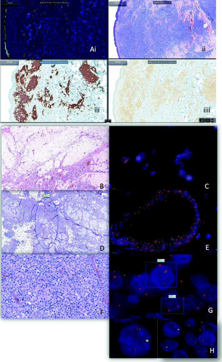
Effective workflow management in a diagnostic pathology laboratory is critical to achieve rapid turnover while maintaining high quality. Fluorescence in situ hybridization analysis (FISH) is the preferred technique for detecting single chromosomal aberrations in diagnostic surgical pathology.

MATERIAL AND METHODS

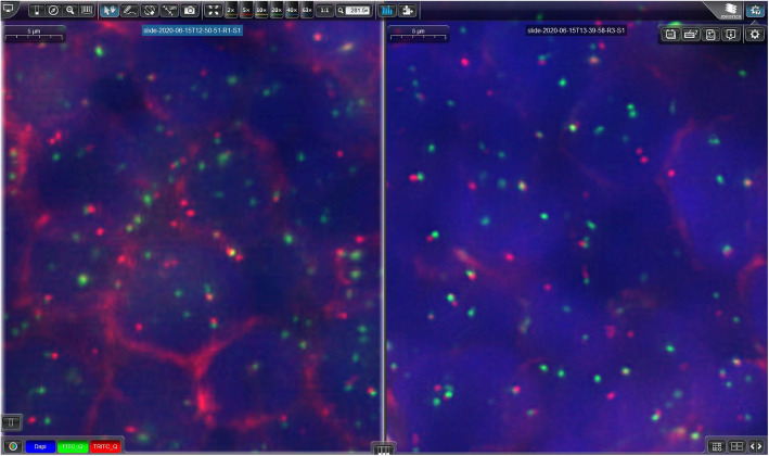
FISH analysis applying a rapid hybridization protocol and using an automated whole-slide fluorescence scanning device (3DHISTECH, Sysmex, Switzerland) were implemented in our workflow. By analyzing 42 diagnostic cases, effects of two different scanning profiles on scanning time, and device memory usage were investigated. Manual signal counting (CaseViewer) and software based signal counting (FISHQuant) were compared.

RESULTS

The two scanning profiles, both including a Z-stack function, differed in their exposure time and digital gain. The "low profile" setting (LP) resulted in a significantly shorter scanning time and lower storage volume compared to the "high profile" (HP) setting, making the LP ideal for routine applications. Both signal counting methods (manual versus software based) provided similar cut-offs on a test-cohort of 13 samples.

CONCLUSION

Scanning FISH slides provides good picture quality, reduces the analysis time and allows easy picture archiving and facilitates remote diagnostics, allowing an effective workflow.

摘要

背景

在诊断病理学实验室中,有效的工作流程管理对于实现快速周转同时保持高质量至关重要。荧光原位杂交分析(FISH)是诊断外科病理学中单条染色体异常检测的首选技术。

材料与方法

我们在工作流程中实施了应用快速杂交方案和自动化全切片荧光扫描设备(3DHISTECH,Sysmex,瑞士)的 FISH 分析。通过分析 42 个诊断病例,研究了两种不同扫描方案对扫描时间和设备内存使用的影响。比较了手动信号计数(CaseViewer)和基于软件的信号计数(FISHQuant)。

结果

两种扫描方案均包含 Z 堆叠功能,但曝光时间和数字增益不同。与“高配置”(HP)设置相比,“低配置”(LP)设置的扫描时间明显更短,存储量更低,因此 LP 非常适合常规应用。两种信号计数方法(手动与基于软件)在 13 个样本的测试队列上提供了相似的截止值。

结论

扫描 FISH 载玻片可提供良好的图像质量,缩短分析时间,并允许轻松进行图像存档和远程诊断,从而实现有效的工作流程。

https://cdn.ncbi.nlm.nih.gov/pmc/blobs/dfa6/8114497/7ea3558539fd/13000_2021_1103_Fig2_HTML.jpg
https://cdn.ncbi.nlm.nih.gov/pmc/blobs/dfa6/8114497/11aaa138e73a/13000_2021_1103_Fig1_HTML.jpg
https://cdn.ncbi.nlm.nih.gov/pmc/blobs/dfa6/8114497/7ea3558539fd/13000_2021_1103_Fig2_HTML.jpg
https://cdn.ncbi.nlm.nih.gov/pmc/blobs/dfa6/8114497/11aaa138e73a/13000_2021_1103_Fig1_HTML.jpg
https://cdn.ncbi.nlm.nih.gov/pmc/blobs/dfa6/8114497/7ea3558539fd/13000_2021_1103_Fig2_HTML.jpg

相似文献

1
Optimized workflow for digitalized FISH analysis in pathology.优化的病理学数字化 FISH 分析工作流程。
Diagn Pathol. 2021 May 11;16(1):42. doi: 10.1186/s13000-021-01103-5.
2
Review of imaging solutions for integrated quantitative immunohistochemistry in the Pathology daily practice.病理学日常实践中集成定量免疫组织化学成像解决方案综述
Folia Histochem Cytobiol. 2009 Jan;47(3):349-54. doi: 10.2478/v10042-008-0114-4.
3
Whole-slide imaging is a robust alternative to traditional fluorescent microscopy for fluorescence in situ hybridization imaging using break-apart DNA probes.全玻片成像技术是一种强大的替代传统荧光显微镜的方法,可用于使用分离 DNA 探针的荧光原位杂交成像。
Hum Pathol. 2013 Aug;44(8):1544-55. doi: 10.1016/j.humpath.2012.12.009. Epub 2013 Mar 18.
4
Digital pathology: current status and future perspectives.数字病理学:现状与未来展望。
Histopathology. 2012 Jul;61(1):1-9. doi: 10.1111/j.1365-2559.2011.03814.x. Epub 2011 Apr 11.
5
Fluorescence in situ hybridization (FISH) signal analysis using automated generated projection images.使用自动生成的投影图像进行荧光原位杂交 (FISH) 信号分析。
Anal Cell Pathol (Amst). 2012;35(5-6):395-405. doi: 10.3233/ACP-2012-0068.
6
Digital pathology: exploring its applications in diagnostic surgical pathology practice.数字病理学:探索其在诊断外科病理学实践中的应用。
Pathology. 2010;42(6):512-8. doi: 10.3109/00313025.2010.508787.
7
iPathology cockpit diagnostic station: validation according to College of American Pathologists Pathology and Laboratory Quality Center recommendation at the Hospital Trust and University of Verona.iPathology 座舱诊断工作站:根据美国病理学家学会病理与实验室质量中心的建议,在维罗纳医院信托和大学进行验证。
Diagn Pathol. 2014;9 Suppl 1(Suppl 1):S12. doi: 10.1186/1746-1596-9-S1-S12. Epub 2014 Dec 19.
8
Applications and challenges of digital pathology and whole slide imaging.数字病理学与全切片成像的应用及挑战
Biotech Histochem. 2015 Jul;90(5):341-7. doi: 10.3109/10520295.2015.1044566. Epub 2015 May 15.
9
Feasibility of using the Omnyx digital pathology system for cytology practice.使用Omnyx数字病理系统进行细胞学实践的可行性。
J Am Soc Cytopathol. 2019 Jul-Aug;8(4):182-189. doi: 10.1016/j.jasc.2019.01.003. Epub 2019 Jan 18.
10
Fast-FISH detection and semi-automated image analysis of numerical chromosome aberrations in hematological malignancies.血液系统恶性肿瘤中染色体数目异常的快速荧光原位杂交检测及半自动图像分析
Anal Cell Pathol. 1998;16(4):211-22. doi: 10.1155/1998/764986.

引用本文的文献

1
Insights into brain tumor diagnosis: exploring hybridization techniques.脑肿瘤诊断的见解:探索杂交技术。
Front Neurol. 2024 Jul 3;15:1393572. doi: 10.3389/fneur.2024.1393572. eCollection 2024.
2
Recurrent and novel fusions detected by targeted RNA sequencing as part of the diagnostic workflow of soft tissue and bone tumours.通过靶向 RNA 测序检测到的复发性和新型融合,作为软组织和骨肿瘤诊断工作流程的一部分。
J Pathol Clin Res. 2024 May;10(3):e12376. doi: 10.1002/2056-4538.12376.
3
Validation of MYC and BCL6 rapid break apart digital fluorescence in situ hybridization assays for clinical use.

本文引用的文献

1
Primary extranodal diffuse large B-cell lymphomas: Many sites, many entities? Clinico-pathological, immunohistochemical and cytogenetic study of 106 cases.原发性结外弥漫性大B细胞淋巴瘤:部位众多,实体各异?106例临床病理、免疫组化及细胞遗传学研究
Cancer Genet. 2018 Dec;228-229:28-40. doi: 10.1016/j.cancergen.2018.08.001. Epub 2018 Aug 28.
2
Fluorescent In Situ Hybridization in Surgical Pathology Practice.荧光原位杂交在外科病理学中的应用。
Adv Anat Pathol. 2018 Jul;25(4):223-237. doi: 10.1097/PAP.0000000000000194.
3
USP6 activation in nodular fasciitis by promoter-swapping gene fusions.
用于临床的MYC和BCL6快速解离数字荧光原位杂交检测的验证
Int J Clin Exp Pathol. 2023 Apr 15;16(4):76-85. eCollection 2023.
结节性筋膜炎中通过启动子交换基因融合激活 USP6。
Mod Pathol. 2017 Nov;30(11):1577-1588. doi: 10.1038/modpathol.2017.78. Epub 2017 Jul 28.
4
Detection of ROS1 rearrangement in non-small cell lung cancer: current and future perspectives.非小细胞肺癌中ROS1重排的检测:现状与未来展望
Lung Cancer (Auckl). 2017 Jul 7;8:45-55. doi: 10.2147/LCTT.S120172. eCollection 2017.
5
Practical use and utility of fluorescence in situ hybridization in the pathological diagnosis of soft tissue and bone tumors.荧光原位杂交技术在软组织和骨肿瘤病理诊断中的实际应用与效用
J Orthop Sci. 2017 Jul;22(4):601-612. doi: 10.1016/j.jos.2017.02.004. Epub 2017 Mar 6.
6
Validation of break-apart and fusion MYC probes using a digital fluorescence in situ hybridization capture and imaging system.使用数字荧光原位杂交捕获和成像系统对断裂-分离和融合MYC探针进行验证。
J Pathol Inform. 2016 May 4;7:20. doi: 10.4103/2153-3539.181764. eCollection 2016.
7
Chromosomal aberrations of cancer-testis antigens in myeloma patients.
Hematol Oncol. 2015 Sep;33(3):159-63. doi: 10.1002/hon.2143. Epub 2014 May 13.
8
Numerical and structural genomic aberrations are reliably detectable in tissue microarrays of formalin-fixed paraffin-embedded tumor samples by fluorescence in-situ hybridization.通过荧光原位杂交技术,可在福尔马林固定石蜡包埋肿瘤样本的组织微阵列中可靠地检测到数值和结构基因组畸变。
PLoS One. 2014 Apr 14;9(4):e95047. doi: 10.1371/journal.pone.0095047. eCollection 2014.
9
Whole-slide imaging is a robust alternative to traditional fluorescent microscopy for fluorescence in situ hybridization imaging using break-apart DNA probes.全玻片成像技术是一种强大的替代传统荧光显微镜的方法,可用于使用分离 DNA 探针的荧光原位杂交成像。
Hum Pathol. 2013 Aug;44(8):1544-55. doi: 10.1016/j.humpath.2012.12.009. Epub 2013 Mar 18.