• 文献检索
  • 文档翻译
  • 深度研究
  • 学术资讯
  • Suppr Zotero 插件Zotero 插件
  • 邀请有礼
  • 套餐&价格
  • 历史记录
应用&插件
Suppr Zotero 插件Zotero 插件浏览器插件Mac 客户端Windows 客户端微信小程序
定价
高级版会员购买积分包购买API积分包
服务
文献检索文档翻译深度研究API 文档MCP 服务
关于我们
关于 Suppr公司介绍联系我们用户协议隐私条款
关注我们

Suppr 超能文献

核心技术专利:CN118964589B侵权必究
粤ICP备2023148730 号-1Suppr @ 2026

文献检索

告别复杂PubMed语法,用中文像聊天一样搜索,搜遍4000万医学文献。AI智能推荐,让科研检索更轻松。

立即免费搜索

文件翻译

保留排版,准确专业,支持PDF/Word/PPT等文件格式,支持 12+语言互译。

免费翻译文档

深度研究

AI帮你快速写综述,25分钟生成高质量综述,智能提取关键信息,辅助科研写作。

立即免费体验

活体介观荧光分子断层成像允许对乳腺癌生长动态进行非侵入性的体内实时监测和定量分析。

Intravital mesoscopic fluorescence molecular tomography allows non-invasive in vivo monitoring and quantification of breast cancer growth dynamics.

机构信息

Cell Biology and Biophysics Unit, European Molecular Biology Laboratory (EMBL), Heidelberg, Germany.

Electrical and Electronics Engineering Department, Karadeniz Technical University, Trabzon, Turkey.

出版信息

Commun Biol. 2021 May 11;4(1):556. doi: 10.1038/s42003-021-02063-8.

DOI:10.1038/s42003-021-02063-8
PMID:33976362
原文链接:https://pmc.ncbi.nlm.nih.gov/articles/PMC8113483/
Abstract

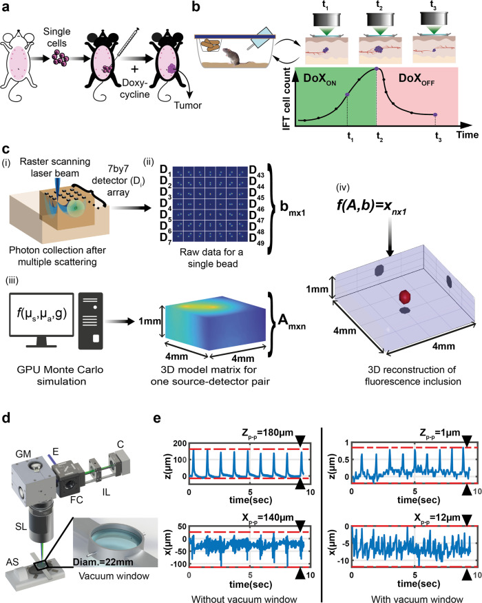
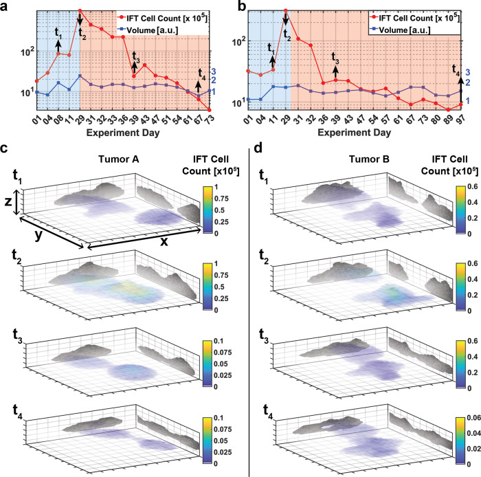
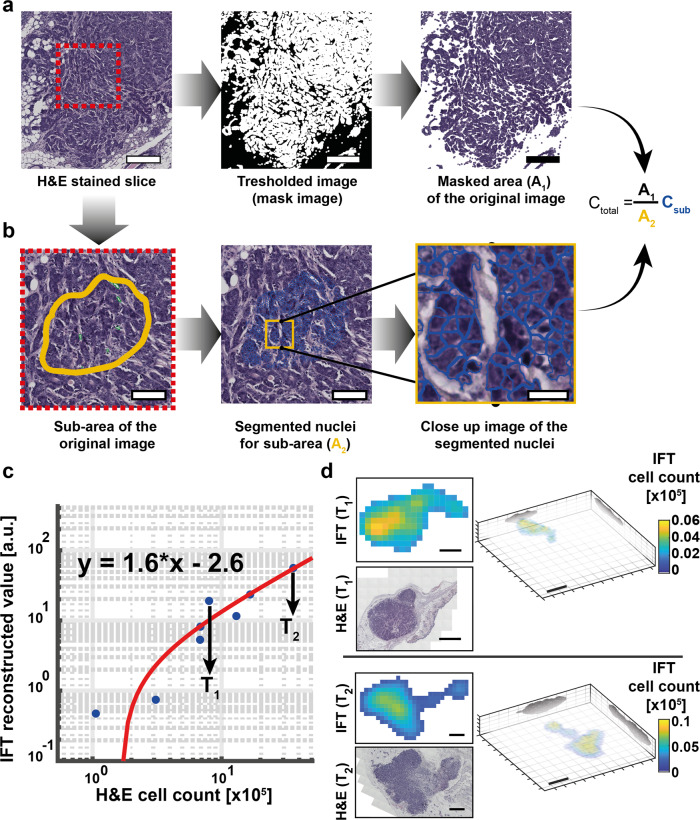
Preclinical breast tumor models are an invaluable tool to systematically study tumor progression and treatment response, yet methods to non-invasively monitor the involved molecular and mechanistic properties under physiologically relevant conditions are limited. Here we present an intravital mesoscopic fluorescence molecular tomography (henceforth IFT) approach that is capable of tracking fluorescently labeled tumor cells in a quantitative manner inside the mammary gland of living mice. Our mesoscopic approach is entirely non-invasive and thus permits prolonged observational periods of several months. The relatively high sensitivity and spatial resolution further enable inferring the overall number of oncogene-expressing tumor cells as well as their tumor volume over the entire cycle from early tumor growth to residual disease following the treatment phase. Our IFT approach is a promising method for studying tumor growth dynamics in a quantitative and longitudinal fashion in-vivo.

摘要

临床前乳腺肿瘤模型是系统研究肿瘤进展和治疗反应的宝贵工具,但在生理相关条件下非侵入性监测所涉及的分子和机制特性的方法有限。在这里,我们提出了一种活体介观荧光分子断层扫描(简称 IFT)方法,该方法能够以定量的方式在活体小鼠的乳腺内跟踪荧光标记的肿瘤细胞。我们的介观方法完全是非侵入性的,因此可以允许长达几个月的长时间观察期。相对较高的灵敏度和空间分辨率还能够推断出整个周期内表达致癌基因的肿瘤细胞的总数,以及在治疗阶段后从早期肿瘤生长到残留疾病的肿瘤体积。我们的 IFT 方法是一种很有前途的方法,可用于在体内以定量和纵向的方式研究肿瘤生长动力学。

https://cdn.ncbi.nlm.nih.gov/pmc/blobs/ff0f/8113483/a60f71931a5a/42003_2021_2063_Fig4_HTML.jpg
https://cdn.ncbi.nlm.nih.gov/pmc/blobs/ff0f/8113483/e8a36e13b039/42003_2021_2063_Fig1_HTML.jpg
https://cdn.ncbi.nlm.nih.gov/pmc/blobs/ff0f/8113483/5760648a1298/42003_2021_2063_Fig2_HTML.jpg
https://cdn.ncbi.nlm.nih.gov/pmc/blobs/ff0f/8113483/5c4302940e7d/42003_2021_2063_Fig3_HTML.jpg
https://cdn.ncbi.nlm.nih.gov/pmc/blobs/ff0f/8113483/a60f71931a5a/42003_2021_2063_Fig4_HTML.jpg

相似文献

1
Intravital mesoscopic fluorescence molecular tomography allows non-invasive in vivo monitoring and quantification of breast cancer growth dynamics.活体介观荧光分子断层成像允许对乳腺癌生长动态进行非侵入性的体内实时监测和定量分析。
Commun Biol. 2021 May 11;4(1):556. doi: 10.1038/s42003-021-02063-8.
2
Simple and Robust Intravital Microscopy Procedures in Hybrid TIE2GFP-BALB/c Transgenic Mice.在杂交 TIE2GFP-BALB/c 转基因小鼠中进行简单而稳健的活体显微镜检查程序。
Mol Imaging Biol. 2020 Jun;22(3):486-493. doi: 10.1007/s11307-019-01442-2.
3
Visualizing murine breast and melanoma tumor microenvironment using intravital multiphoton microscopy.利用活体多光子显微镜观察小鼠乳腺和黑色素瘤肿瘤微环境。
STAR Protoc. 2021 Aug 19;2(3):100722. doi: 10.1016/j.xpro.2021.100722. eCollection 2021 Sep 17.
4
Longitudinal live animal micro-CT allows for quantitative analysis of tumor-induced bone destruction.纵向活体小动物 micro-CT 可用于定量分析肿瘤诱导的骨破坏。
Bone. 2011 Jan;48(1):141-51. doi: 10.1016/j.bone.2010.05.033. Epub 2010 Jun 1.
5
An Intravital Microscopy Toolbox to Study Mammary Gland Dynamics from Cellular Level to Organ Scale.一种用于从细胞水平到器官尺度研究乳腺动态的活体显微镜工具包。
J Mammary Gland Biol Neoplasia. 2021 Mar;26(1):9-27. doi: 10.1007/s10911-021-09487-2. Epub 2021 May 4.
6
Intravital imaging reveals systemic ezrin inhibition impedes cancer cell migration and lymph node metastasis in breast cancer.活体成像显示,系统抑制埃兹蛋白可阻碍乳腺癌细胞迁移和淋巴结转移。
Breast Cancer Res. 2019 Jan 24;21(1):12. doi: 10.1186/s13058-018-1079-7.
7
Two Photon Intravital Microscopy of Lyme Borrelia in Mice.小鼠中莱姆病螺旋体的双光子活体显微镜检查
Methods Mol Biol. 2018;1690:279-290. doi: 10.1007/978-1-4939-7383-5_20.
8
Metaboloptics: Visualization of the tumor functional landscape via metabolic and vascular imaging.代谢光学:通过代谢和血管成像可视化肿瘤功能景观。
Sci Rep. 2018 Mar 8;8(1):4171. doi: 10.1038/s41598-018-22480-w.
9
Biodistribution Analysis of NIR-Labeled Nanogels Using in Vivo FMT Imaging in Triple Negative Human Mammary Carcinoma Models.近红外标记纳米凝胶的体内 FMT 成像生物分布分析在三阴性人乳腺癌模型中的应用。
Mol Pharm. 2018 Mar 5;15(3):1180-1191. doi: 10.1021/acs.molpharmaceut.7b01011. Epub 2018 Feb 12.
10
Preparation and Testing of Cells Expressing Fluorescent Proteins for Intravital Imaging of Tumor Microenvironment.用于肿瘤微环境活体成像的荧光蛋白表达细胞的制备与检测
Bull Exp Biol Med. 2019 May;167(1):123-130. doi: 10.1007/s10517-019-04475-3. Epub 2019 Jun 10.

引用本文的文献

1
FROP-1 peptide-conjugated ultrasmall superparamagnetic nanoparticles as a targeted T1-weighted MR contrast agent for breast cancer: in vitro study.FROP-1肽偶联的超小超顺磁性纳米颗粒作为乳腺癌的靶向T1加权磁共振造影剂:体外研究
BMC Biomed Eng. 2025 May 1;7(1):5. doi: 10.1186/s42490-025-00091-7.
2
Imagining the future of optical microscopy: everything, everywhere, all at once.想象光学显微镜的未来:一切、无处不在、一切都在瞬间。
Commun Biol. 2023 Oct 28;6(1):1096. doi: 10.1038/s42003-023-05468-9.
3
active-targeting fluorescence molecular imaging with adaptive background fluorescence subtraction.

本文引用的文献

1
High-resolution tomographic analysis of in vitro 3D glioblastoma tumor model under long-term drug treatment.高分辨率断层分析在体外 3D 脑胶质瘤肿瘤模型中长期药物处理下的表现。
Sci Adv. 2020 Mar 6;6(10):eaay7513. doi: 10.1126/sciadv.aay7513. eCollection 2020 Mar.
2
Graphics processing unit-accelerated mesh-based Monte Carlo photon transport simulations.基于图形处理单元加速的网格蒙特卡罗光子传输模拟。
J Biomed Opt. 2019 Nov;24(11):1-6. doi: 10.1117/1.JBO.24.11.115002.
3
Ductal carcinoma in situ: to treat or not to treat, that is the question.
具有自适应背景荧光扣除的主动靶向荧光分子成像
Front Oncol. 2023 Mar 14;13:1130155. doi: 10.3389/fonc.2023.1130155. eCollection 2023.
4
Design and characterization of a time-domain optical tomography platform for mesoscopic lifetime imaging.用于介观寿命成像的时域光学层析成像平台的设计与特性分析
Biomed Opt Express. 2022 Aug 10;13(9):4637-4651. doi: 10.1364/BOE.460216. eCollection 2022 Sep 1.
5
Multidimensional Imaging of Breast Cancer.乳腺癌的多维成像。
Cold Spring Harb Perspect Med. 2023 May 2;13(5):a041330. doi: 10.1101/cshperspect.a041330.
6
3D k-space reflectance fluorescence tomography via deep learning.基于深度学习的 3D k-空间反射荧光层析成像。
Opt Lett. 2022 Mar 15;47(6):1533-1536. doi: 10.1364/OL.450935.
导管原位癌:治疗还是不治疗,这是个问题。
Br J Cancer. 2019 Aug;121(4):285-292. doi: 10.1038/s41416-019-0478-6. Epub 2019 Jul 9.
4
Improving mesoscopic fluorescence molecular tomography via preconditioning and regularization.通过预处理和正则化改进介观荧光分子断层扫描技术。
Biomed Opt Express. 2018 May 23;9(6):2765-2778. doi: 10.1364/BOE.9.002765. eCollection 2018 Jun 1.
5
Imaging stem cell distribution, growth, migration, and differentiation in 3-D scaffolds for bone tissue engineering using mesoscopic fluorescence tomography.使用介观荧光层析成像技术在 3D 支架中对用于骨组织工程的干细胞进行分布、生长、迁移和分化的成像。
Biotechnol Bioeng. 2018 Jan;115(1):257-265. doi: 10.1002/bit.26452.
6
Improving mesoscopic fluorescence molecular tomography through data reduction.通过数据约简改进介观荧光分子断层成像
Biomed Opt Express. 2017 Jul 28;8(8):3868-3881. doi: 10.1364/BOE.8.003868. eCollection 2017 Aug 1.
7
Predicting the response to CTLA-4 blockade by longitudinal noninvasive monitoring of CD8 T cells.通过对CD8 T细胞进行纵向无创监测来预测对CTLA-4阻断的反应。
J Exp Med. 2017 Aug 7;214(8):2243-2255. doi: 10.1084/jem.20161950. Epub 2017 Jun 30.
8
Metabolic shifts in residual breast cancer drive tumor recurrence.残留乳腺癌中的代谢转变驱动肿瘤复发。
J Clin Invest. 2017 Jun 1;127(6):2091-2105. doi: 10.1172/JCI89914. Epub 2017 May 15.
9
Imaging windows for long-term intravital imaging: General overview and technical insights.用于长期活体成像的成像窗口:概述与技术见解
Intravital. 2014 Aug 11;3(2):e29917. doi: 10.4161/intv.29917. eCollection 2014.
10
Genetically engineered mouse models in oncology research and cancer medicine.肿瘤学研究与癌症医学中的基因工程小鼠模型
EMBO Mol Med. 2017 Feb;9(2):137-153. doi: 10.15252/emmm.201606857.