Sun Jinju, Liu Kaijun, Tong Haipeng, Liu Huan, Li Xiaoguang, Luo Yi, Li Yang, Yao Yun, Jin Rongbing, Fang Jingqin, Chen Xiao
Department of Nuclear Medicine, Daping Hospital, Army Medical University, Chongqing, China.
Department of Gastroenterology, Daping Hospital, Army Medical University, Chongqing, China.
Front Oncol. 2021 Apr 26;11:634564. doi: 10.3389/fonc.2021.634564. eCollection 2021.
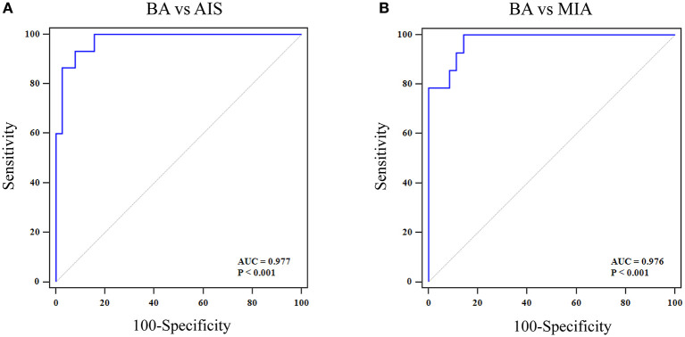
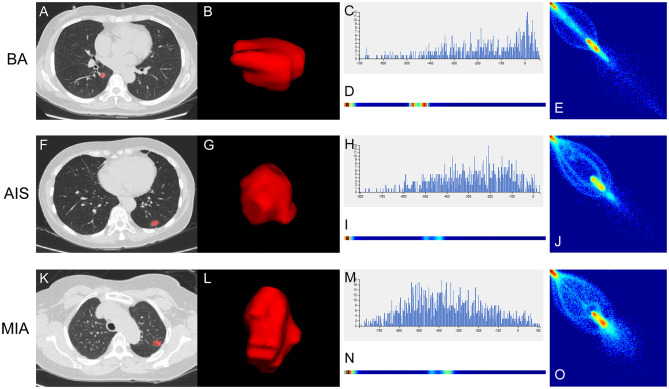
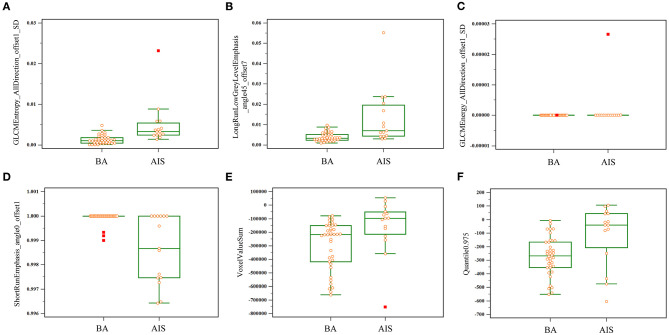
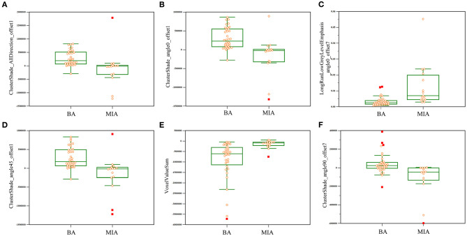
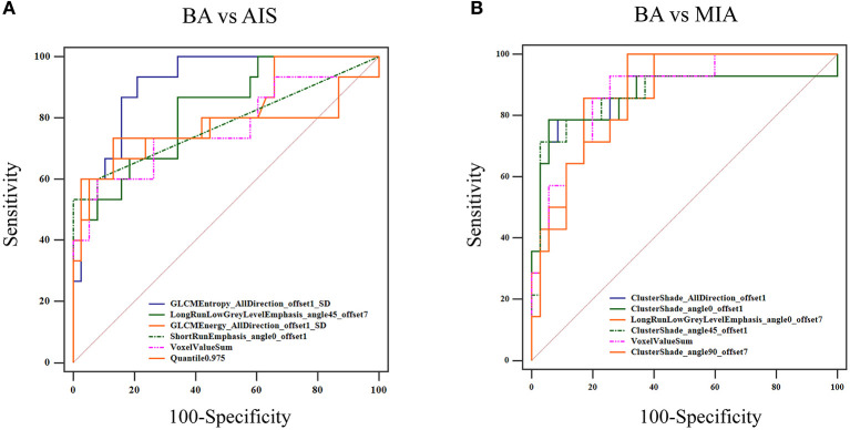
This study aimed to investigate the potential of computed tomography (CT) imaging features and texture analysis to distinguish bronchiolar adenoma (BA) from adenocarcinoma (AIS)/minimally invasive adenocarcinoma (MIA). Fifteen patients with BA, 38 patients with AIS, and 36 patients with MIA were included in this study. Clinical data and CT imaging features of the three lesions were evaluated. Texture features were extracted from the thin-section unenhanced CT images using Artificial Intelligence Kit software. Then, multivariate logistic regression analysis based on selected texture features was employed to distinguish BA from AIS/MIA. Receiver operating characteristics curves were performed to determine the diagnostic performance of the features. By comparison with AIS/MIA, significantly different CT imaging features of BA included nodule type, tumor size, and pseudo-cavitation sign. Among them, pseudo-cavitation sign had a moderate diagnostic value for distinguishing BA and AIS/MIA (AUC: 0.741 and 0.708, respectively). Further, a total of 396 quantitative texture features were extracted. After comparation, the top six texture features showing the most significant difference between BA and AIS or MIA were chosen. The ROC results showed that these key texture features had a high diagnostic value for differentiating BA from AIS or MIA, among which the value of a comprehensive model with six selected texture features was the highest (AUC: 0.977 or 0.976, respectively) for BA and AIS or MIA. These results indicated that texture analyses can effectively improve the efficacy of thin-section unenhanced CT for discriminating BA from AIS/MIA. CT texture analysis can effectively improve the efficacy of thin-section unenhanced CT for discriminating BA from AIS/MIA, which has a potential clinical value and helps pathologist and clinicians to make diagnostic and therapeutic strategies.
本研究旨在探讨计算机断层扫描(CT)成像特征和纹理分析在区分细支气管腺瘤(BA)与原位腺癌(AIS)/微浸润腺癌(MIA)方面的潜力。本研究纳入了15例BA患者、38例AIS患者和36例MIA患者。评估了这三种病变的临床资料和CT成像特征。使用人工智能套件软件从薄层平扫CT图像中提取纹理特征。然后,基于选定的纹理特征进行多因素逻辑回归分析,以区分BA与AIS/MIA。绘制受试者工作特征曲线以确定这些特征的诊断性能。与AIS/MIA相比,BA具有显著差异的CT成像特征包括结节类型、肿瘤大小和假空洞征。其中,假空洞征在区分BA与AIS/MIA方面具有中等诊断价值(AUC分别为0.741和0.708)。此外,共提取了396个定量纹理特征。经过比较,选择了BA与AIS或MIA之间差异最显著的前六个纹理特征。ROC结果显示,这些关键纹理特征在区分BA与AIS或MIA方面具有较高的诊断价值,其中包含六个选定纹理特征的综合模型对BA与AIS或MIA的诊断价值最高(AUC分别为0.977或0.976)。这些结果表明,纹理分析可有效提高薄层平扫CT区分BA与AIS/MIA的效能。CT纹理分析可有效提高薄层平扫CT区分BA与AIS/MIA的效能,具有潜在的临床价值,有助于病理学家和临床医生制定诊断和治疗策略。