Ribbat-Idel Julika, Dressler Franz F, Krupar Rosemarie, Watermann Christian, Paulsen Finn-Ole, Kuppler Patrick, Klapper Luise, Offermann Anne, Wollenberg Barbara, Rades Dirk, Laban Simon, Reischl Markus, Bruchhage Karl-Ludwig, Idel Christian, Perner Sven
Institute of Pathology, University of Luebeck and University Hospital Schleswig-Holstein, Luebeck, Germany.
Pathology, Research Center Borstel, Leibniz Lung Center, Borstel, Germany.
Front Med (Lausanne). 2021 Apr 27;8:640515. doi: 10.3389/fmed.2021.640515. eCollection 2021.
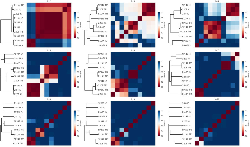
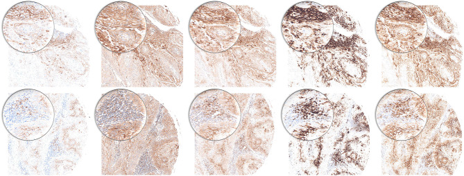
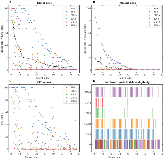
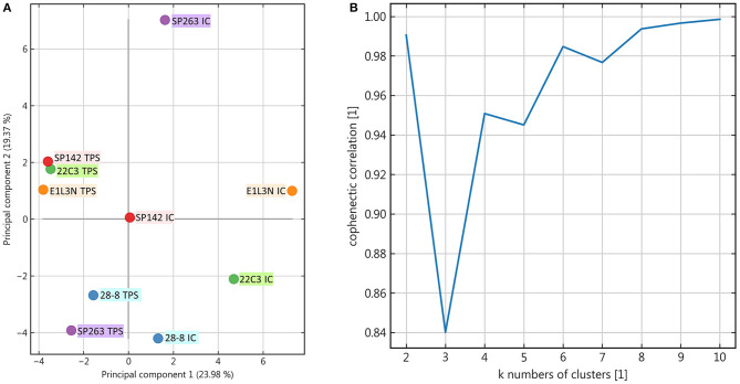
The approval of immune checkpoint inhibitors in combination with specific diagnostic biomarkers presents new challenges to pathologists as tumor tissue needs to be tested for expression of programmed death-ligand 1 (PD-L1) for a variety of indications. As there is currently no requirement to use companion diagnostic assays for PD-L1 testing in Germany different clones are used in daily routine. While the correlation of staining results has been tested in various entities, there is no data for head and neck squamous cell carcinomas (HNSCC) so far. We tested five different PD-L1 clones (SP263, SP142, E1L3N, 22-8, 22C3) on primary HNSCC tumor tissue of 75 patients in the form of tissue microarrays. Stainings of both immune and tumor cells were then assessed and quantified by pathologists to simulate real-world routine diagnostics. The results were analyzed descriptively and the resulting staining pattern across patients was further investigated by principal component analysis and non-negative matrix factorization clustering. Percentages of positive immune and tumor cells varied greatly. Both the resulting combined positive score as well as the eligibility for certain checkpoint inhibitor regimens was therefore strongly dependent on the choice of the antibody. No relevant co-clustering and low similarity of relative staining patterns across patients was found for the different antibodies. Performance of different diagnostic anti PD-L1 antibody clones in HNSCC is less robust and interchangeable compared to reported data from other tumor entities. Determination of PD-L1 expression is critical for therapeutic decision making and may be aided by back-to-back testing of different PD-L1 clones.
免疫检查点抑制剂与特定诊断生物标志物联合使用的获批给病理学家带来了新挑战,因为针对多种适应症,需要检测肿瘤组织中程序性死亡配体1(PD-L1)的表达。由于德国目前对PD-L1检测不要求使用配套诊断检测方法,日常工作中使用了不同的克隆体。虽然在各种实体中已对染色结果的相关性进行了测试,但到目前为止,尚无关于头颈部鳞状细胞癌(HNSCC)的数据。我们以组织微阵列的形式,对75例患者的原发性HNSCC肿瘤组织测试了五种不同的PD-L1克隆体(SP263、SP142、E1L3N、22-8、22C3)。然后由病理学家对免疫细胞和肿瘤细胞的染色进行评估和定量,以模拟实际临床常规诊断。对结果进行描述性分析,并通过主成分分析和非负矩阵分解聚类进一步研究患者的染色模式。免疫细胞和肿瘤细胞阳性百分比差异很大。因此,最终的综合阳性评分以及某些检查点抑制剂治疗方案的适用性在很大程度上取决于抗体的选择。不同抗体在患者中未发现相关的共聚类,相对染色模式的相似性较低。与其他肿瘤实体报告的数据相比,不同诊断性抗PD-L1抗体克隆体在HNSCC中的性能较差,且不可互换。PD-L1表达的测定对于治疗决策至关重要,对不同PD-L1克隆体进行背对背检测可能会有所帮助。