CÚRAM SFI Research Centre for Medical Devices, National University of Ireland, Galway, Ireland.
CEA, CNRS, MIRCen, Laboratoire des Maladies Neurodégénératives, Université Paris-Saclay, Fontenay-aux-Roses, France.
J Neuroinflammation. 2021 May 16;18(1):116. doi: 10.1186/s12974-021-02163-6.
Neuroinflammation is an underlying pathology of all neurological conditions, the understanding of which is still being comprehended. A specific molecular pathway that has been overlooked in neuroinflammation is glycosylation (i.e., post-translational addition of glycans to the protein structure). N-glycosylation is a specific type of glycosylation with a cardinal role in the central nervous system (CNS), which is highlighted by congenital glycosylation diseases that result in neuropathological symptoms such as epilepsy and mental retardation. Changes in N-glycosylation can ultimately affect glycoproteins' functions, which will have an impact on cell machinery. Therefore, characterisation of N-glycosylation alterations in a neuroinflammatory scenario can provide a potential target for future therapies.
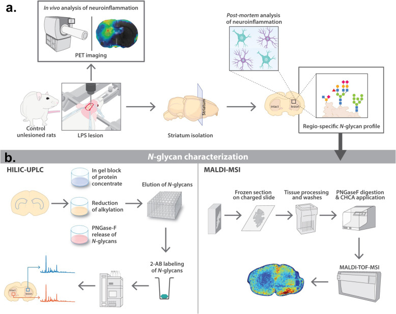
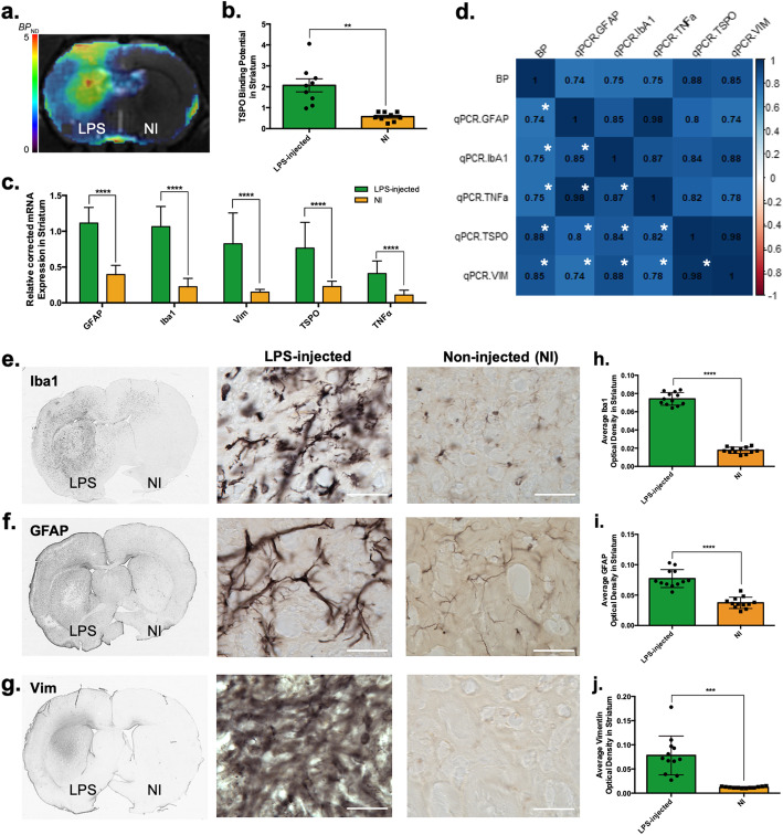
With that aim, the unilateral intrastriatal injection of lipopolysaccharide (LPS) in the adult rat brain was used as a model of neuroinflammation. In vivo and post-mortem, quantitative and spatial characterisation of both neuroinflammation and N-glycome was performed at 1-week post-injection of LPS. These aspects were investigated through a multifaceted approach based on positron emission tomography (PET), quantitative histology, reverse transcription-quantitative polymerase chain reaction (RT-qPCR), liquid chromatography and matrix-assisted laser desorption ionisation mass spectrometry imaging (MALDI-MSI).
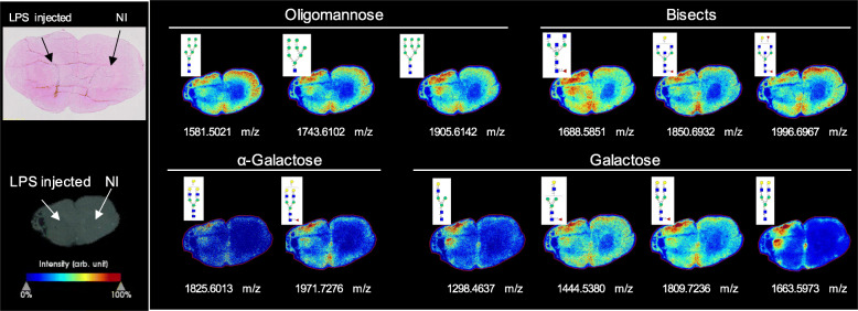
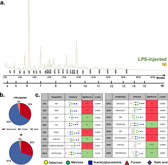
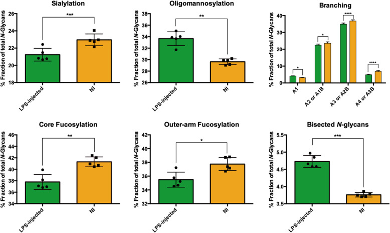
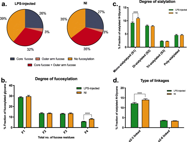
In the brain region showing LPS-induced neuroinflammation, a significant decrease in the abundance of sialylated and core fucosylated structures was seen (approximately 7.5% and 8.5%, respectively), whereas oligomannose N-glycans were significantly increased (13.5%). This was confirmed by MALDI-MSI, which provided a high-resolution spatial distribution of N-glycans, allowing precise comparison between normal and diseased brain hemispheres.
Together, our data show for the first time the complete profiling of N-glycomic changes in a well-characterised animal model of neuroinflammation. These data represent a pioneering step to identify critical targets that may modulate neuroinflammation in neurodegenerative diseases.
神经炎症是所有神经疾病的潜在病理学,对其的理解仍在进行中。在神经炎症中,一个被忽视的特定分子途径是糖基化(即糖链在后翻译阶段添加到蛋白质结构中)。N-糖基化是糖基化的一种特殊类型,在中枢神经系统(CNS)中起着至关重要的作用,这一点在导致神经病理学症状(如癫痫和智力迟钝)的先天性糖基化疾病中得到了强调。N-糖基化的变化最终会影响糖蛋白的功能,从而影响细胞机制。因此,在神经炎症情况下对 N-糖基化变化的特征描述可以为未来的治疗提供潜在的靶点。
为此,我们使用成年大鼠大脑单侧纹状体注射脂多糖(LPS)作为神经炎症模型。在体内和死后,通过基于正电子发射断层扫描(PET)、定量组织学、反转录定量聚合酶链反应(RT-qPCR)、液相色谱和基质辅助激光解吸电离质谱成像(MALDI-MSI)的多方面方法,对 LPS 注射后 1 周的神经炎症和 N-聚糖组进行了定量和空间特征描述。
在表现出 LPS 诱导的神经炎症的大脑区域,发现唾液酸化和核心岩藻糖化结构的丰度显著降低(分别约为 7.5%和 8.5%),而寡甘露糖 N-聚糖显著增加(13.5%)。MALDI-MSI 证实了这一点,它提供了 N-聚糖的高分辨率空间分布,允许在正常和患病大脑半球之间进行精确比较。
总之,我们的数据首次全面描绘了神经炎症的特征性动物模型中 N-糖组的变化。这些数据代表了确定可能调节神经退行性疾病中神经炎症的关键靶点的开创性步骤。