Kushibar Kaisar, Salem Mostafa, Valverde Sergi, Rovira Àlex, Salvi Joaquim, Oliver Arnau, Lladó Xavier
Institute of Computer Vision and Robotics, University of Girona, Girona, Spain.
Computer Science Department, Faculty of Computers and Information, Assiut University, Asyut, Egypt.
Front Neurosci. 2021 Apr 29;15:608808. doi: 10.3389/fnins.2021.608808. eCollection 2021.
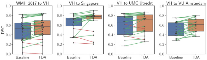
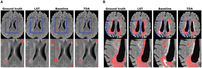
Segmentation of brain images from Magnetic Resonance Images (MRI) is an indispensable step in clinical practice. Morphological changes of sub-cortical brain structures and quantification of brain lesions are considered biomarkers of neurological and neurodegenerative disorders and used for diagnosis, treatment planning, and monitoring disease progression. In recent years, deep learning methods showed an outstanding performance in medical image segmentation. However, these methods suffer from generalisability problem due to inter-centre and inter-scanner variabilities of the MRI images. The main objective of the study is to develop an automated deep learning segmentation approach that is accurate and robust to the variabilities in scanner and acquisition protocols. In this paper, we propose a transductive transfer learning approach for domain adaptation to reduce the domain-shift effect in brain MRI segmentation. The transductive scenario assumes that there are sets of images from two different domains: (1) source-images with manually annotated labels; and (2) target-images without expert annotations. Then, the network is jointly optimised integrating both source and target images into the transductive training process to segment the regions of interest and to minimise the domain-shift effect. We proposed to use a histogram loss in the feature level to carry out the latter optimisation problem. In order to demonstrate the benefit of the proposed approach, the method has been tested in two different brain MRI image segmentation problems using multi-centre and multi-scanner databases for: (1) sub-cortical brain structure segmentation; and (2) white matter hyperintensities segmentation. The experiments showed that the segmentation performance of a pre-trained model could be significantly improved by up to 10%. For the first segmentation problem it was possible to achieve a maximum improvement from 0.680 to 0.799 in average Dice Similarity Coefficient (DSC) metric and for the second problem the average DSC improved from 0.504 to 0.602. Moreover, the improvements after domain adaptation were on par or showed better performance compared to the commonly used traditional unsupervised segmentation methods (FIRST and LST), also achieving faster execution time. Taking this into account, this work presents one more step toward the practical implementation of deep learning algorithms into the clinical routine.
从磁共振成像(MRI)中分割脑图像是临床实践中不可或缺的一步。皮质下脑结构的形态变化和脑病变的量化被视为神经和神经退行性疾病的生物标志物,并用于诊断、治疗规划和监测疾病进展。近年来,深度学习方法在医学图像分割中表现出卓越的性能。然而,由于MRI图像的中心间和扫描仪间的变异性,这些方法存在泛化问题。本研究的主要目标是开发一种自动化的深度学习分割方法,该方法对扫描仪和采集协议中的变异性准确且稳健。在本文中,我们提出一种用于域适应的转导迁移学习方法,以减少脑MRI分割中的域转移效应。转导场景假设存在来自两个不同域的图像集:(1)带有手动标注标签的源图像;以及(2)没有专家注释的目标图像。然后,将源图像和目标图像都集成到转导训练过程中,对网络进行联合优化,以分割感兴趣区域并最小化域转移效应。我们建议在特征级别使用直方图损失来解决后一个优化问题。为了证明所提出方法的益处,该方法已在两个不同的脑MRI图像分割问题中进行了测试,使用多中心和多扫描仪数据库用于:(1)皮质下脑结构分割;以及(2)白质高信号分割。实验表明,预训练模型的分割性能可显著提高多达10%。对于第一个分割问题,平均骰子相似系数(DSC)指标从0.680最大提高到0.799,对于第二个问题,平均DSC从0.504提高到0.602。此外,与常用的传统无监督分割方法(FIRST和LST)相比,域适应后的改进相当或表现更好,执行时间也更快。考虑到这一点,这项工作朝着将深度学习算法实际应用于临床常规又迈进了一步。