• 文献检索
  • 文档翻译
  • 深度研究
  • 学术资讯
  • Suppr Zotero 插件Zotero 插件
  • 邀请有礼
  • 套餐&价格
  • 历史记录
应用&插件
Suppr Zotero 插件Zotero 插件浏览器插件Mac 客户端Windows 客户端微信小程序
定价
高级版会员购买积分包购买API积分包
服务
文献检索文档翻译深度研究API 文档MCP 服务
关于我们
关于 Suppr公司介绍联系我们用户协议隐私条款
关注我们

Suppr 超能文献

核心技术专利:CN118964589B侵权必究
粤ICP备2023148730 号-1Suppr @ 2026

文献检索

告别复杂PubMed语法,用中文像聊天一样搜索,搜遍4000万医学文献。AI智能推荐,让科研检索更轻松。

立即免费搜索

文件翻译

保留排版,准确专业,支持PDF/Word/PPT等文件格式,支持 12+语言互译。

免费翻译文档

深度研究

AI帮你快速写综述,25分钟生成高质量综述,智能提取关键信息,辅助科研写作。

立即免费体验

通过首次通过灌注心血管磁共振对全定量心肌血流图进行自动节段分析。

Automated Segmental Analysis of Fully Quantitative Myocardial Blood Flow Maps by First-Pass Perfusion Cardiovascular Magnetic Resonance.

作者信息

Jacobs Matthew, Benovoy Mitchel, Chang Lin-Ching, Corcoran David, Berry Colin, Arai Andrew E, Hsu Li-Yueh

机构信息

National Heart, Lung, and Blood Institute, National Institutes of Health, Bethesda, MD 20892, USA.

Department of Electrical Engineering and Computer Science, The Catholic University of America, Washington, DC 20064, USA.

出版信息

IEEE Access. 2021;9:52796-52811. doi: 10.1109/access.2021.3070320. Epub 2021 Apr 1.

DOI:10.1109/access.2021.3070320
PMID:33996344
原文链接:https://pmc.ncbi.nlm.nih.gov/articles/PMC8117952/
Abstract

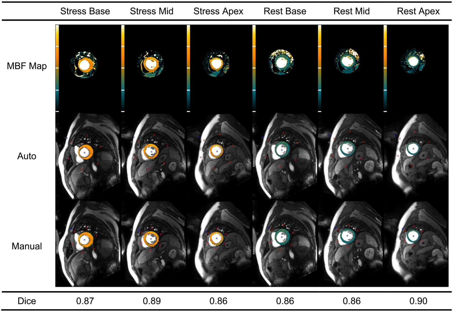
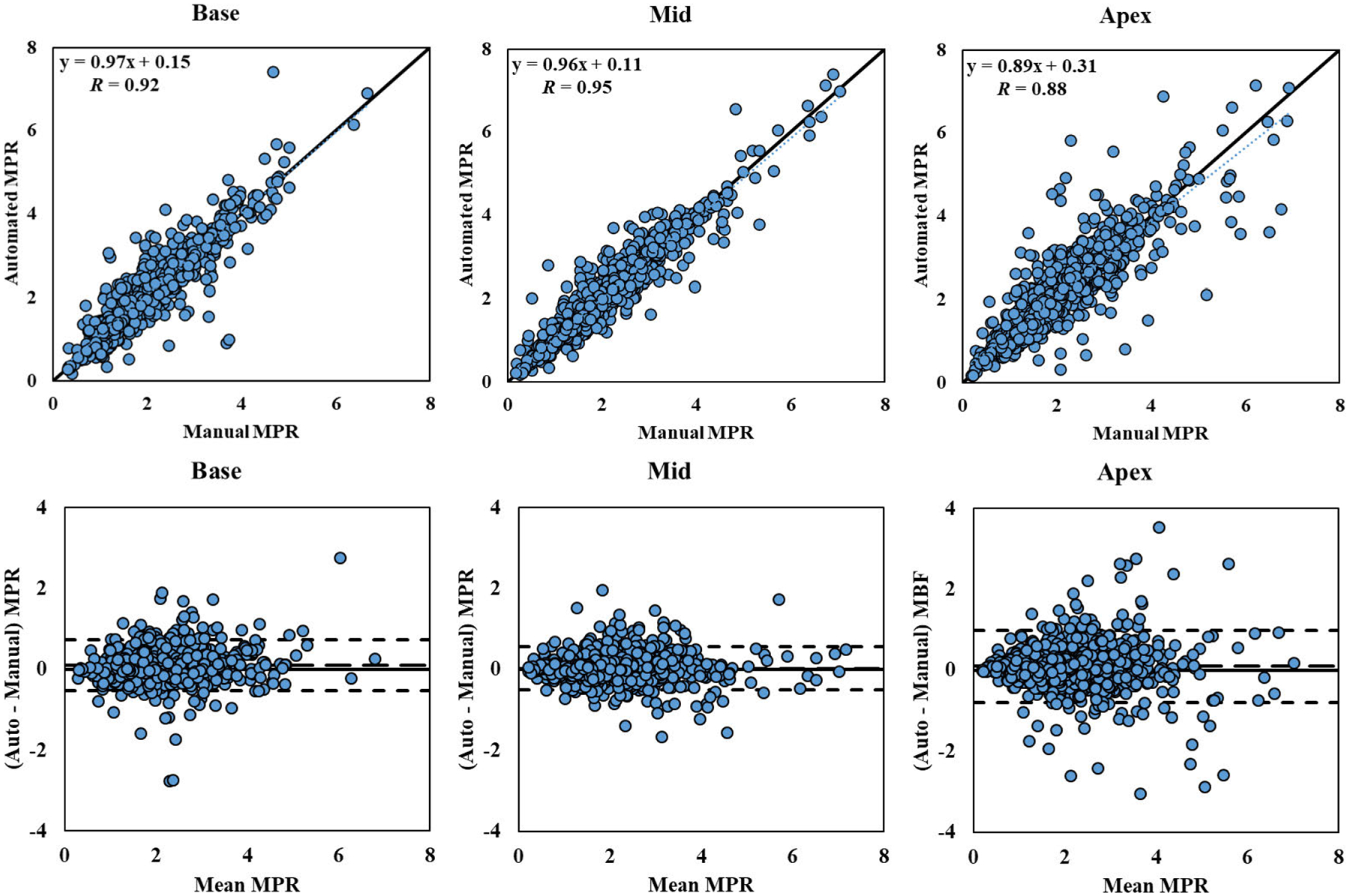
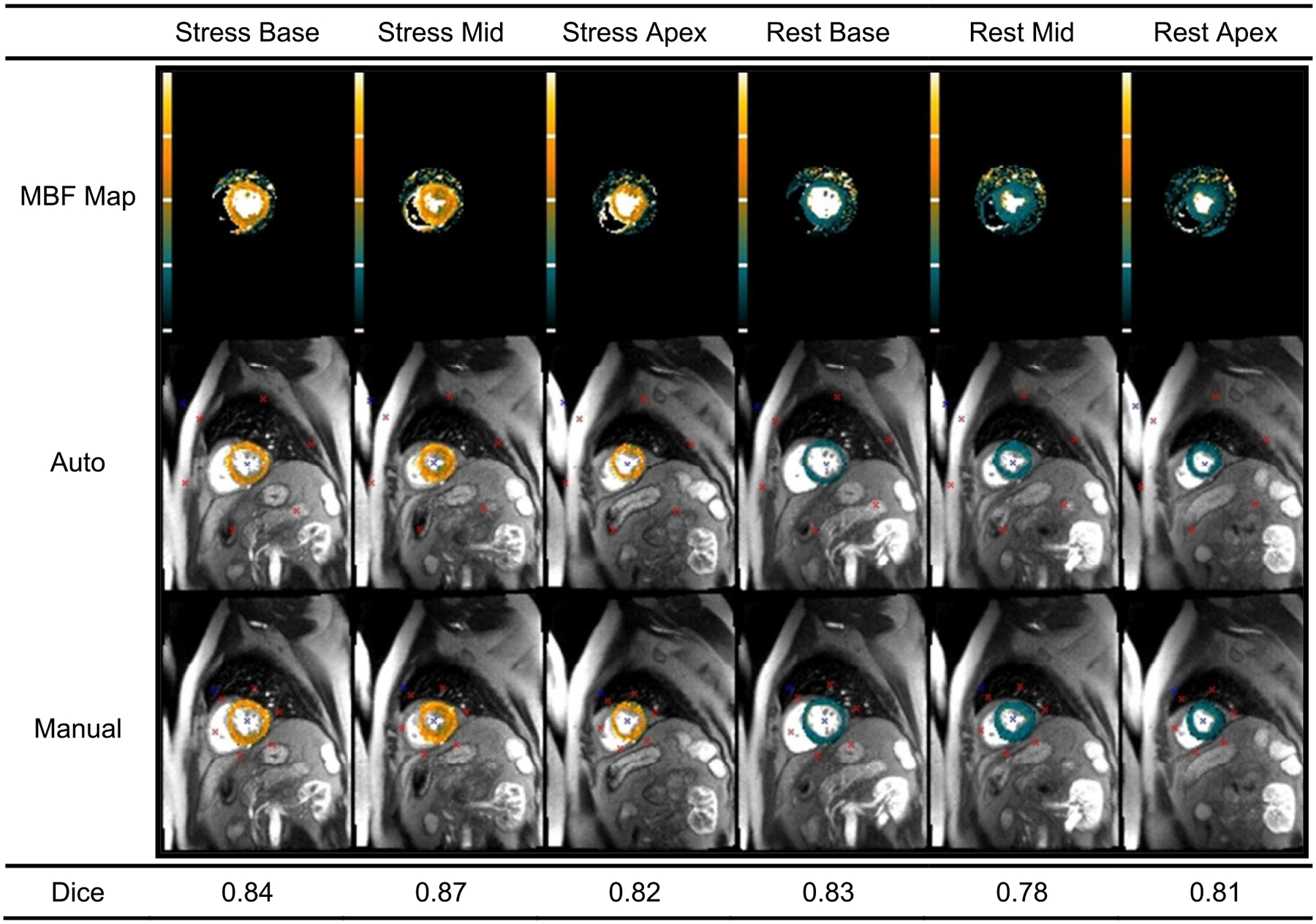
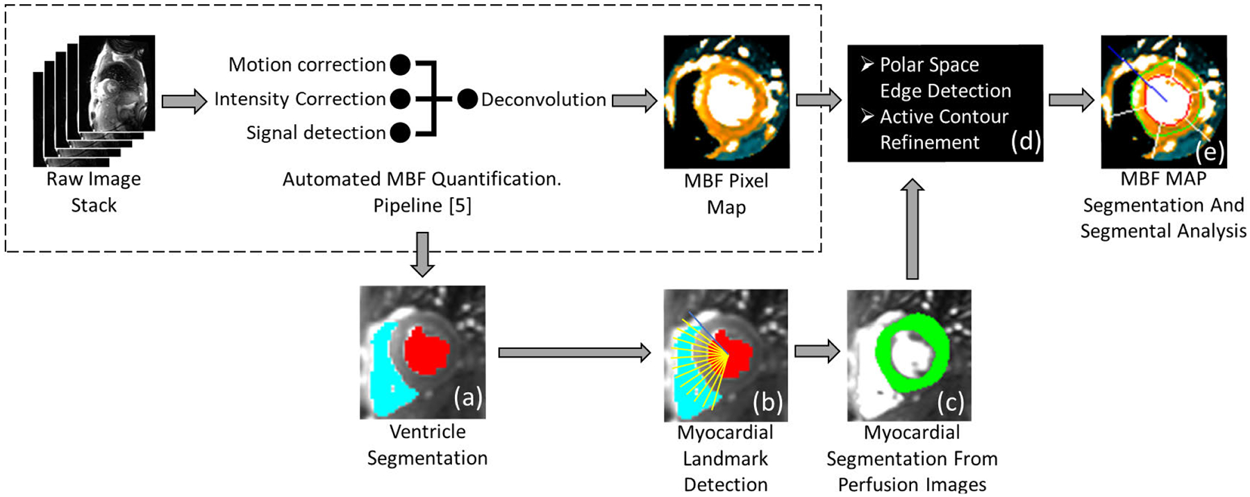
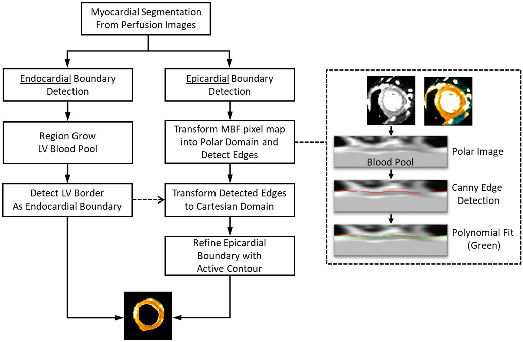
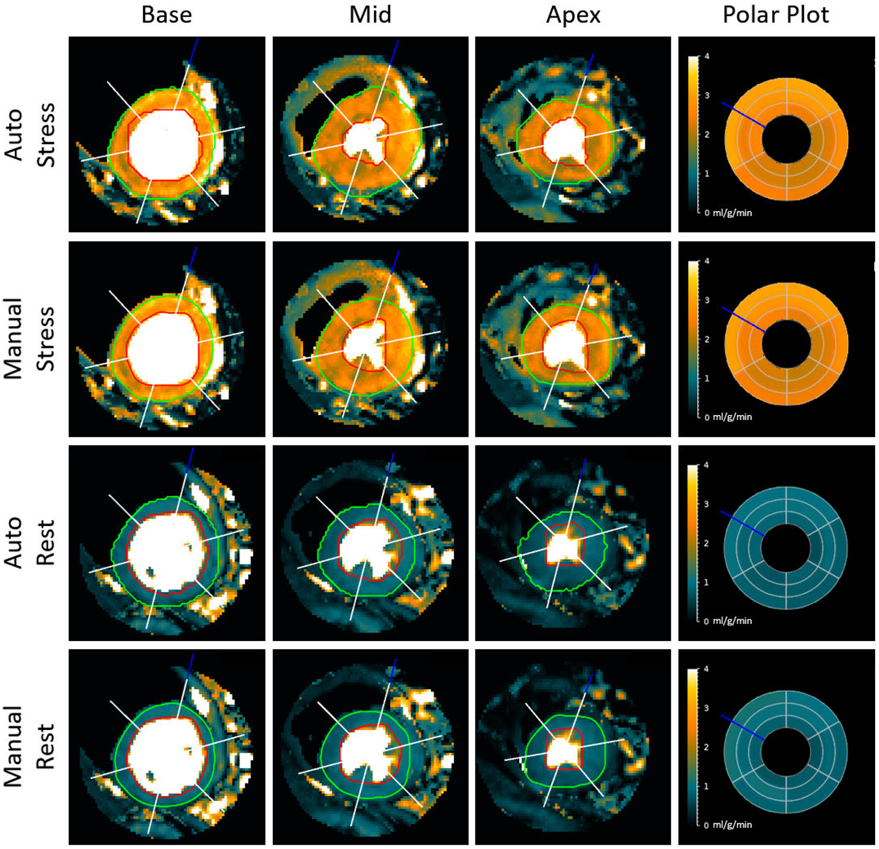
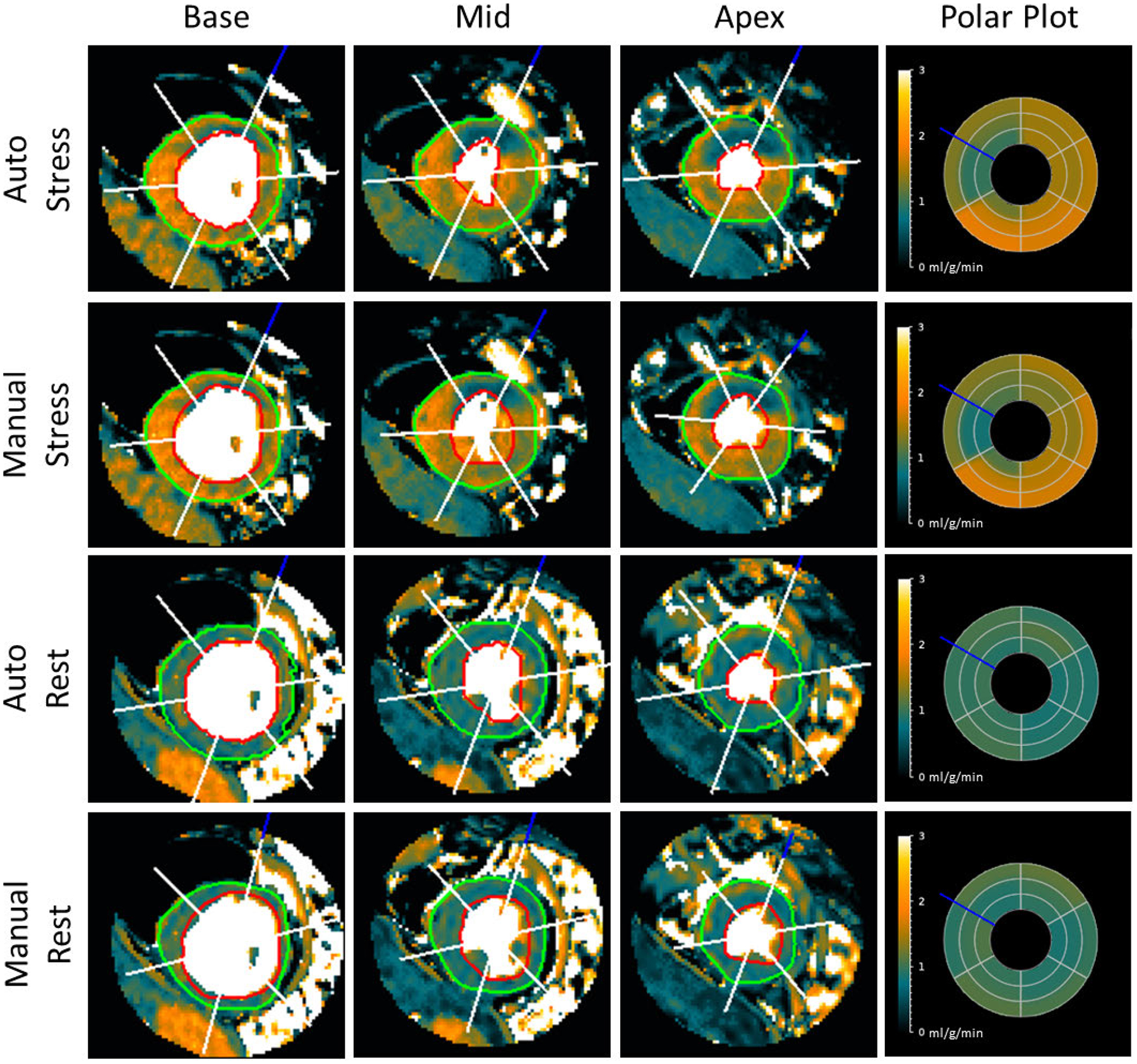
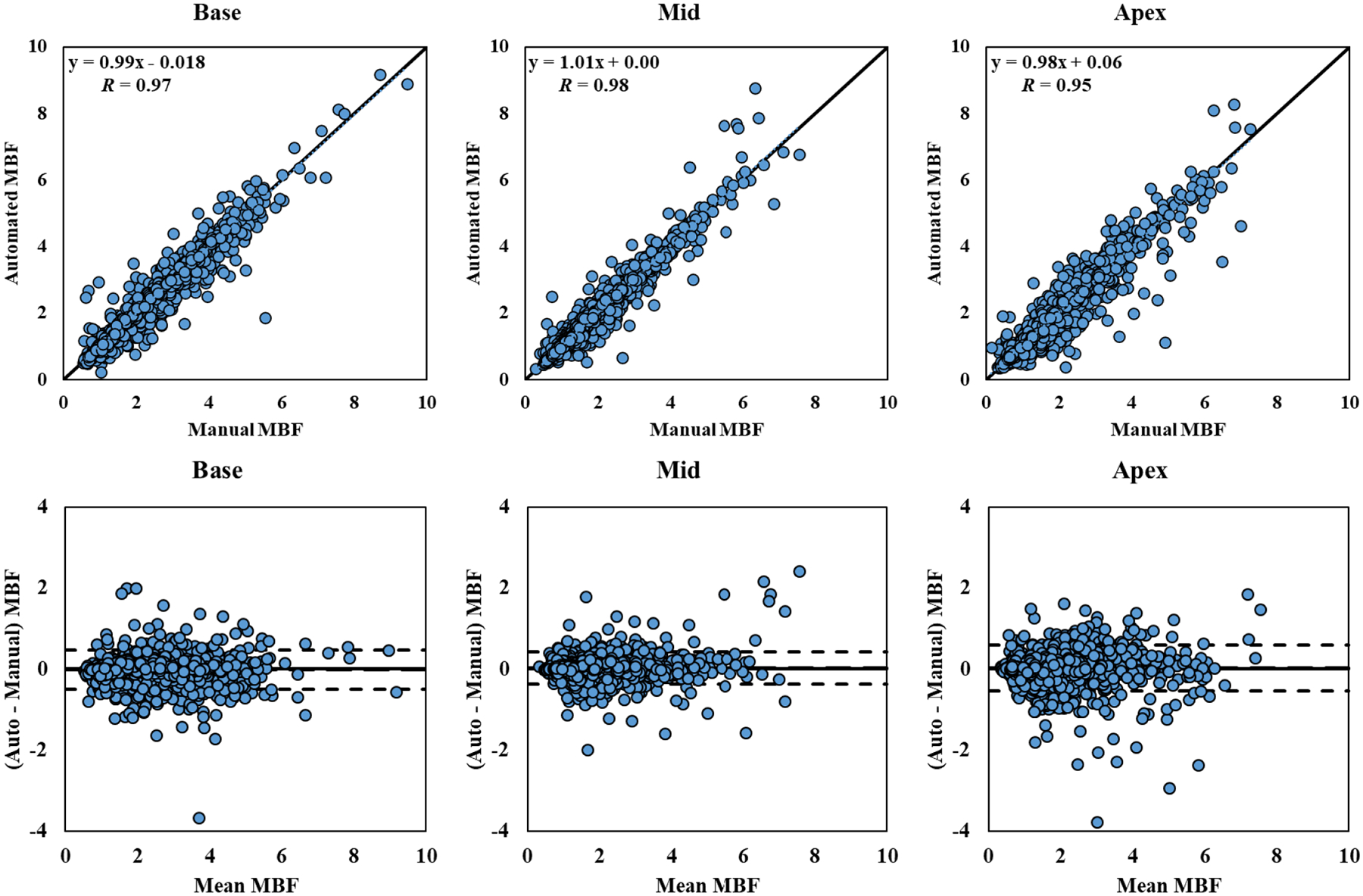
First pass gadolinium-enhanced cardiovascular magnetic resonance (CMR) perfusion imaging allows fully quantitative pixel-wise myocardial blood flow (MBF) assessment, with proven diagnostic value for coronary artery disease. Segmental analysis requires manual segmentation of the myocardium. This work presents a fully automatic method of segmenting the left ventricular myocardium from MBF pixel maps, validated on a retrospective dataset of 247 clinical CMR perfusion studies, each including rest and stress images of three slice locations, performed on a 1.5T scanner. Pixel-wise MBF maps were segmented using an automated pipeline including region growing, edge detection, principal component analysis, and active contours to segment the myocardium, detect key landmarks, and divide the myocardium into sectors appropriate for analysis. Automated segmentation results were compared against a manually defined reference standard using three quantitative metrics: Dice coefficient, Cohen Kappa and myocardial border distance. Sector-wise average MBF and myocardial perfusion reserve (MPR) were compared using Pearson's correlation coefficient and Bland-Altman Plots. The proposed method segmented stress and rest MBF maps of 243 studies automatically. Automated and manual myocardial segmentation had an average (± standard deviation) Dice coefficient of 0.86 ± 0.06, Cohen Kappa of 0.86 ± 0.06, and Euclidian distances of 1.47 ± 0.73 mm and 1.02 ± 0.51 mm for the epicardial and endocardial border, respectively. Automated and manual sector-wise MBF and MPR values correlated with Pearson's coefficient of 0.97 and 0.92, respectively, while Bland-Altman analysis showed bias of 0.01 and 0.07 ml/g/min. The validated method has been integrated with our fully automated MBF pixel mapping pipeline to aid quantitative assessment of myocardial perfusion CMR.

摘要

首次通过钆增强心血管磁共振(CMR)灌注成像可实现全定量逐像素心肌血流(MBF)评估,对冠状动脉疾病具有已证实的诊断价值。节段分析需要对心肌进行手动分割。这项工作提出了一种从MBF像素图中自动分割左心室心肌的方法,并在247例临床CMR灌注研究的回顾性数据集中进行了验证,每个数据集包括在1.5T扫描仪上对三个切片位置进行的静息和负荷图像。使用包括区域生长、边缘检测、主成分分析和活动轮廓的自动化流程对逐像素MBF图进行分割,以分割心肌、检测关键地标,并将心肌划分为适合分析的扇区。使用三个定量指标将自动分割结果与手动定义的参考标准进行比较:骰子系数、科恩卡帕系数和心肌边界距离。使用皮尔逊相关系数和布兰德-奥特曼图比较扇区平均MBF和心肌灌注储备(MPR)。所提出的方法自动分割了243项研究的负荷和静息MBF图。自动和手动心肌分割的平均(±标准差)骰子系数为0.86±0.06,科恩卡帕系数为0.86±0.06,心外膜和心内膜边界的欧几里得距离分别为1.47±0.73mm和1.02±0.51mm。自动和手动扇区MBF及MPR值的皮尔逊相关系数分别为0.97和0.92,而布兰德-奥特曼分析显示偏差分别为0.01和0.07ml/g/min。该经过验证的方法已与我们的全自动MBF像素映射流程集成,以辅助心肌灌注CMR的定量评估。

https://cdn.ncbi.nlm.nih.gov/pmc/blobs/e9d8/8117952/017753565a83/nihms-1692921-f0014.jpg
https://cdn.ncbi.nlm.nih.gov/pmc/blobs/e9d8/8117952/74be6fe86865/nihms-1692921-f0015.jpg
https://cdn.ncbi.nlm.nih.gov/pmc/blobs/e9d8/8117952/f1732f9b5c6f/nihms-1692921-f0016.jpg
https://cdn.ncbi.nlm.nih.gov/pmc/blobs/e9d8/8117952/ba026ca8dafe/nihms-1692921-f0008.jpg
https://cdn.ncbi.nlm.nih.gov/pmc/blobs/e9d8/8117952/9a3f1b7714ce/nihms-1692921-f0009.jpg
https://cdn.ncbi.nlm.nih.gov/pmc/blobs/e9d8/8117952/2c1c6d643f09/nihms-1692921-f0010.jpg
https://cdn.ncbi.nlm.nih.gov/pmc/blobs/e9d8/8117952/451047c6d35b/nihms-1692921-f0011.jpg
https://cdn.ncbi.nlm.nih.gov/pmc/blobs/e9d8/8117952/6e99d0ca6186/nihms-1692921-f0012.jpg
https://cdn.ncbi.nlm.nih.gov/pmc/blobs/e9d8/8117952/00bc84fe2297/nihms-1692921-f0013.jpg
https://cdn.ncbi.nlm.nih.gov/pmc/blobs/e9d8/8117952/017753565a83/nihms-1692921-f0014.jpg

相似文献

1
Automated Segmental Analysis of Fully Quantitative Myocardial Blood Flow Maps by First-Pass Perfusion Cardiovascular Magnetic Resonance.通过首次通过灌注心血管磁共振对全定量心肌血流图进行自动节段分析。
IEEE Access. 2021;9:52796-52811. doi: 10.1109/access.2021.3070320. Epub 2021 Apr 1.
2
Fully automated pixel-wise quantitative CMR-myocardial perfusion with CMR-coronary angiography to detect hemodynamically significant coronary artery disease.应用 CMR 冠状动脉造影全自动像素级定量 CMR 心肌灌注成像检测有血流动力学意义的冠状动脉疾病。
Eur Radiol. 2023 Oct;33(10):7238-7249. doi: 10.1007/s00330-023-09689-8. Epub 2023 May 5.
3
Diagnostic Performance of Fully Automated Pixel-Wise Quantitative Myocardial Perfusion Imaging by Cardiovascular Magnetic Resonance.心血管磁共振全自动像素定量心肌灌注成像的诊断性能。
JACC Cardiovasc Imaging. 2018 May;11(5):697-707. doi: 10.1016/j.jcmg.2018.01.005. Epub 2018 Feb 14.
4
A quantitative pixel-wise measurement of myocardial blood flow by contrast-enhanced first-pass CMR perfusion imaging: microsphere validation in dogs and feasibility study in humans.对比增强首过 CMR 灌注成像定量像素级心肌血流测量:微球在犬中的验证和在人体中的可行性研究。
JACC Cardiovasc Imaging. 2012 Feb;5(2):154-66. doi: 10.1016/j.jcmg.2011.07.013.
5
Sex- and age-specific normal values for automated quantitative pixel-wise myocardial perfusion cardiovascular magnetic resonance.基于自动化定量像素心肌灌注心血管磁共振的性别和年龄特异性正常值。
Eur Heart J Cardiovasc Imaging. 2023 Mar 21;24(4):426-434. doi: 10.1093/ehjci/jeac231.
6
Automated Pixel-Wise Quantitative Myocardial Perfusion Mapping by CMR to Detect Obstructive Coronary Artery Disease and Coronary Microvascular Dysfunction: Validation Against Invasive Coronary Physiology.CMR 自动像素定量心肌灌注成像检测冠状动脉疾病和冠状动脉微血管功能障碍:与有创性冠状动脉生理学的对照验证。
JACC Cardiovasc Imaging. 2019 Oct;12(10):1958-1969. doi: 10.1016/j.jcmg.2018.12.022. Epub 2019 Feb 13.
7
Evaluation of an automated method for arterial input function detection for first-pass myocardial perfusion cardiovascular magnetic resonance.用于首次通过心肌灌注心血管磁共振的动脉输入函数检测自动化方法的评估。
J Cardiovasc Magn Reson. 2016 Apr 8;18:17. doi: 10.1186/s12968-016-0239-0.
8
Fully automated, inline quantification of myocardial blood flow with cardiovascular magnetic resonance: repeatability of measurements in healthy subjects.使用心血管磁共振进行全自动、在线心肌血流定量:健康受试者测量的可重复性。
J Cardiovasc Magn Reson. 2018 Jul 9;20(1):48. doi: 10.1186/s12968-018-0462-y.
9
Comparison of dual-bolus versus dual-sequence techniques for determining myocardial blood flow and myocardial perfusion reserve by cardiac magnetic resonance stress perfusion: From the Automated Quantitative analysis of myocardial perfusion cardiac Magnetic Resonance Consortium.心脏磁共振应力灌注中双团注与双序列技术测定心肌血流和心肌灌注储备的比较:来自心肌灌注心脏磁共振自动定量分析联盟。
J Cardiovasc Magn Reson. 2024;26(2):101085. doi: 10.1016/j.jocmr.2024.101085. Epub 2024 Aug 16.
10
High-resolution free-breathing automated quantitative myocardial perfusion by cardiovascular magnetic resonance for the detection of functionally significant coronary artery disease.心血管磁共振高分辨率自由呼吸自动定量心肌灌注成像检测有功能意义的冠状动脉疾病。
Eur Heart J Cardiovasc Imaging. 2024 Jun 28;25(7):914-925. doi: 10.1093/ehjci/jeae084.

引用本文的文献

1
Residual myocardial hyperemia in regadenoson stress/rest quantitative perfusion cardiac magnetic resonance.雷加昔布负荷/静息定量灌注心脏磁共振成像中的残余心肌充血
Radiol Med. 2025 Aug 23. doi: 10.1007/s11547-025-02062-3.
2
Advanced Cardiac Magnetic Resonance Imaging for Assessment of Obstructive Coronary Artery Disease - ADVOCATE-CMR Study Rationale and Design.用于评估阻塞性冠状动脉疾病的高级心脏磁共振成像——ADVOCATE-CMR研究原理与设计
J Cardiovasc Magn Reson. 2025 Apr 25:101900. doi: 10.1016/j.jocmr.2025.101900.
3
Stress T1 mapping and quantitative perfusion cardiovascular magnetic resonance in patients with suspected obstructive coronary artery disease.

本文引用的文献

1
Deep learning approach for the segmentation of aneurysmal ascending aorta.用于升主动脉瘤分割的深度学习方法。
Biomed Eng Lett. 2020 Nov 20;11(1):15-24. doi: 10.1007/s13534-020-00179-0. eCollection 2021 Feb.
2
Automated Inline Analysis of Myocardial Perfusion MRI with Deep Learning.基于深度学习的心肌灌注磁共振成像自动在线分析
Radiol Artif Intell. 2020 Oct 21;2(6):e200009. doi: 10.1148/ryai.2020200009. eCollection 2020 Oct.
3
Deep-Learning-Based Preprocessing for Quantitative Myocardial Perfusion MRI.基于深度学习的定量心肌灌注磁共振成像预处理
疑似阻塞性冠状动脉疾病患者的应力T1映射和定量灌注心血管磁共振成像
Eur Heart J Cardiovasc Imaging. 2025 May 30;26(6):980-990. doi: 10.1093/ehjci/jeaf059.
4
Improved Robustness for Deep Learning-based Segmentation of Multi-Center Myocardial Perfusion MRI Datasets Using Data Adaptive Uncertainty-guided Space-time Analysis.使用数据自适应不确定性引导的时空分析提高基于深度学习的多中心心肌灌注MRI数据集分割的鲁棒性
ArXiv. 2024 Aug 9:arXiv:2408.04805v1.
5
Clinical implementation of a fully automated quantitative perfusion cardiovascular magnetic resonance imaging workflow with a simplified dual-bolus contrast administration scheme.临床应用全自动定量灌注心血管磁共振成像工作流程,采用简化的双对比剂给药方案。
Sci Rep. 2024 Apr 26;14(1):9665. doi: 10.1038/s41598-024-60503-x.
6
High-resolution quantification of stress perfusion defects by cardiac magnetic resonance.通过心脏磁共振对压力灌注缺损进行高分辨率定量分析。
Eur Heart J Imaging Methods Pract. 2024 Jan 9;2(1):qyae001. doi: 10.1093/ehjimp/qyae001. eCollection 2024 Jan.
7
Automatic calculation of myocardial perfusion reserve using deep learning with uncertainty quantification.使用具有不确定性量化的深度学习自动计算心肌灌注储备。
Quant Imaging Med Surg. 2023 Dec 1;13(12):7936-7949. doi: 10.21037/qims-23-840. Epub 2023 Oct 10.
8
Role of pulmonary perfusion magnetic resonance imaging for the diagnosis of pulmonary hypertension: A review.肺灌注磁共振成像在肺动脉高压诊断中的作用:综述
World J Radiol. 2023 Sep 28;15(9):256-273. doi: 10.4329/wjr.v15.i9.256.
9
Focal and diffuse myocardial fibrosis both contribute to regional hypoperfusion assessed by post-processing quantitative-perfusion MRI techniques.局灶性和弥漫性心肌纤维化均会导致通过后处理定量灌注磁共振成像技术评估的局部灌注不足。
Front Cardiovasc Med. 2023 Sep 19;10:1260156. doi: 10.3389/fcvm.2023.1260156. eCollection 2023.
10
Evolving Management Paradigm for Stable Ischemic Heart Disease Patients: JACC Review Topic of the Week.稳定型缺血性心脏病患者管理模式的演变:JACC 本周综述主题。
J Am Coll Cardiol. 2023 Feb 7;81(5):505-514. doi: 10.1016/j.jacc.2022.08.814.
J Magn Reson Imaging. 2020 Jun;51(6):1689-1696. doi: 10.1002/jmri.26983. Epub 2019 Nov 11.
4
Automated Pixel-Wise Quantitative Myocardial Perfusion Mapping by CMR to Detect Obstructive Coronary Artery Disease and Coronary Microvascular Dysfunction: Validation Against Invasive Coronary Physiology.CMR 自动像素定量心肌灌注成像检测冠状动脉疾病和冠状动脉微血管功能障碍:与有创性冠状动脉生理学的对照验证。
JACC Cardiovasc Imaging. 2019 Oct;12(10):1958-1969. doi: 10.1016/j.jcmg.2018.12.022. Epub 2019 Feb 13.
5
Quantitative myocardial perfusion in coronary artery disease: A perfusion mapping study.冠状动脉疾病中的定量心肌灌注:一项灌注绘图研究。
J Magn Reson Imaging. 2019 Sep;50(3):756-762. doi: 10.1002/jmri.26668. Epub 2019 Jan 25.
6
Fully automated, inline quantification of myocardial blood flow with cardiovascular magnetic resonance: repeatability of measurements in healthy subjects.使用心血管磁共振进行全自动、在线心肌血流定量:健康受试者测量的可重复性。
J Cardiovasc Magn Reson. 2018 Jul 9;20(1):48. doi: 10.1186/s12968-018-0462-y.
7
Quantitative Myocardial Perfusion Imaging Versus Visual Analysis in Diagnosing Myocardial Ischemia: A CE-MARC Substudy.定量心肌灌注成像与视觉分析在诊断心肌缺血中的比较:CE-MARC 子研究。
JACC Cardiovasc Imaging. 2018 May;11(5):711-718. doi: 10.1016/j.jcmg.2018.02.019.
8
Diagnostic Performance of Fully Automated Pixel-Wise Quantitative Myocardial Perfusion Imaging by Cardiovascular Magnetic Resonance.心血管磁共振全自动像素定量心肌灌注成像的诊断性能。
JACC Cardiovasc Imaging. 2018 May;11(5):697-707. doi: 10.1016/j.jcmg.2018.01.005. Epub 2018 Feb 14.
9
Prognostic Value of Quantitative Stress Perfusion Cardiac Magnetic Resonance.定量压力灌注心脏磁共振的预后价值
JACC Cardiovasc Imaging. 2018 May;11(5):686-694. doi: 10.1016/j.jcmg.2017.07.022. Epub 2017 Nov 15.
10
Fully quantitative cardiovascular magnetic resonance myocardial perfusion ready for clinical use: a comparison between cardiovascular magnetic resonance imaging and positron emission tomography.全面定量心血管磁共振心肌灌注成像准备临床应用:心血管磁共振成像与正电子发射断层扫描比较。
J Cardiovasc Magn Reson. 2017 Oct 19;19(1):78. doi: 10.1186/s12968-017-0388-9.