• 文献检索
  • 文档翻译
  • 深度研究
  • 学术资讯
  • Suppr Zotero 插件Zotero 插件
  • 邀请有礼
  • 套餐&价格
  • 历史记录
应用&插件
Suppr Zotero 插件Zotero 插件浏览器插件Mac 客户端Windows 客户端微信小程序
定价
高级版会员购买积分包购买API积分包
服务
文献检索文档翻译深度研究API 文档MCP 服务
关于我们
关于 Suppr公司介绍联系我们用户协议隐私条款
关注我们

Suppr 超能文献

核心技术专利:CN118964589B侵权必究
粤ICP备2023148730 号-1Suppr @ 2026

文献检索

告别复杂PubMed语法,用中文像聊天一样搜索,搜遍4000万医学文献。AI智能推荐,让科研检索更轻松。

立即免费搜索

文件翻译

保留排版,准确专业,支持PDF/Word/PPT等文件格式,支持 12+语言互译。

免费翻译文档

深度研究

AI帮你快速写综述,25分钟生成高质量综述,智能提取关键信息,辅助科研写作。

立即免费体验

全内置半月板修复装置结构与拉出缝线用于关节镜下经胫骨修复后内侧半月板根部撕裂的生物力学比较:一项配对尸体研究

Biomechanical Comparison of an All-Inside Meniscal Repair Device Construct Versus Pullout Sutures for Arthroscopic Transtibial Repair of Posterior Medial Meniscus Root Tears: A Matched-Pair Cadaveric Study.

作者信息

Vertullo Christopher J, Cadman Joseph, Dabirrahmani Danè, Appleyard Richard

机构信息

Knee Research Australia and the Centre for Musculoskeletal Research, Menzies Health Institute, Griffith University, Gold Coast, Australia.

Orthopaedic Biomechanics Research Group, Department of Biomedical Sciences, Faculty of Medicine and Health Sciences, Macquarie University, Sydney, Australia.

出版信息

Orthop J Sports Med. 2021 Apr 22;9(4):23259671211000464. doi: 10.1177/23259671211000464. eCollection 2021 Apr.

DOI:10.1177/23259671211000464
PMID:33997064
原文链接:https://pmc.ncbi.nlm.nih.gov/articles/PMC8072104/
Abstract

BACKGROUND

Meniscus root repairs are important for restoring knee function after a complete meniscus root tear. Various suturing patterns have been proposed for the root repair. The 2-simple-stitches (TSS) method is currently the preferred technique, as it is simplest to perform and allows the least displacement of the meniscus root.

PURPOSE

To compare the biomechanical properties of a posterior medial meniscus transtibial root repair consisting of an all-inside meniscal repair device (AMRD) construct with the TSS pullout suture pattern.

STUDY DESIGN

Controlled laboratory study.

METHODS

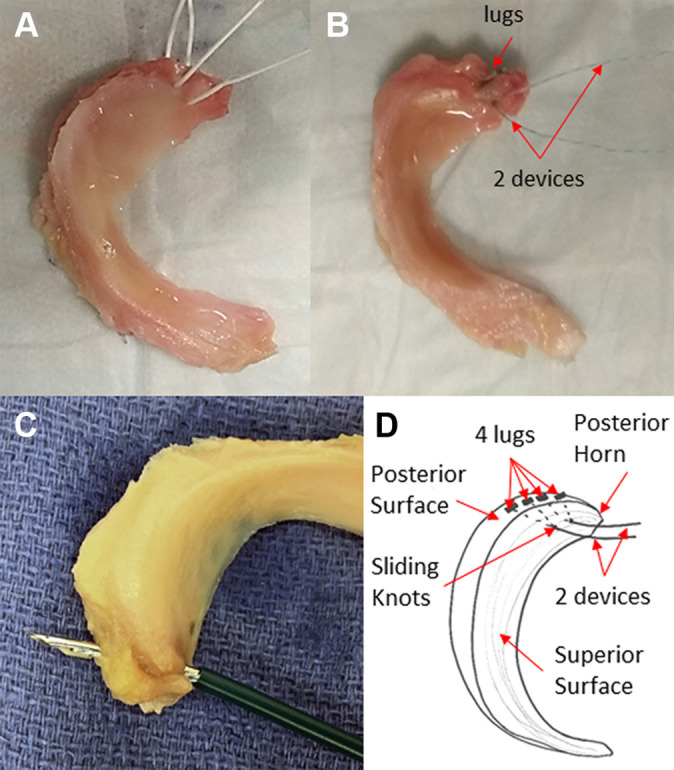
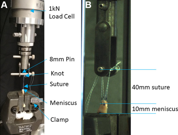
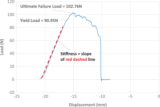
Ten pairs of cadaveric medial menisci were prepared with 1 of the 2 constructs. The constructs were randomized between pairs. All constructs were subjected to preloading with 2 N for 10 seconds and then cyclic loading from 5 N to 20 N for 1000 cycles at a frequency of 0.5 Hz. Subsequently, the menisci were loaded to failure at a rate of 0.5 mm/s. All loads were applied in-line with the circumferential meniscal fibers near the posterior medial meniscal horn.

RESULTS

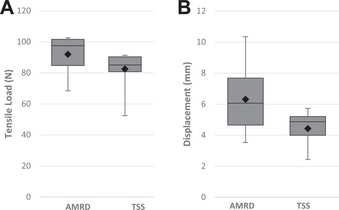
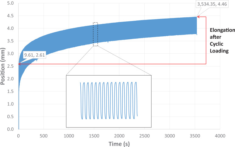
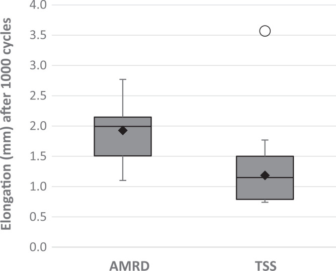
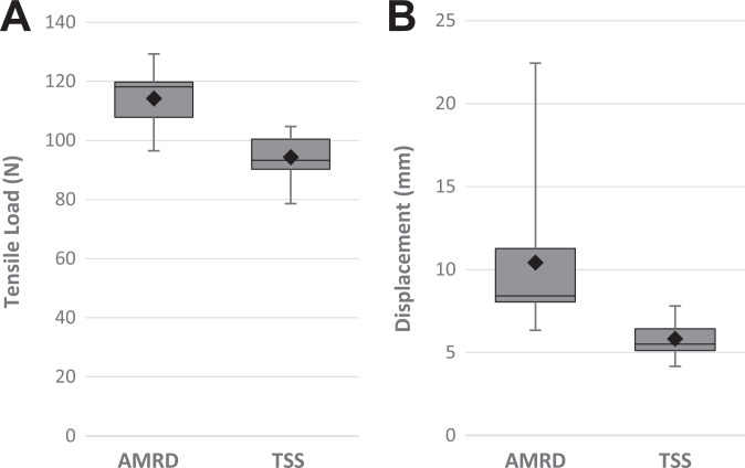
The mean yield load and stiffness were similar for both constructs. The elongation after cyclic loading was greater for the AMRD. The displacement at both yield load and ultimate failure were also higher for the AMRD. The ultimate failure load of the AMRD was also significantly higher. During load to failure, the mode of failure in the AMRD was heterogeneous. All the TSS constructs failed by suture cutout.

CONCLUSION

Posterior medial meniscus root repairs using both the AMRD and TSS constructs have elongation under the biomechanically acceptable threshold of 3 mm. The stiffness and yield loads indicate similar mechanical properties of the constructs. However, the significantly higher elongation for the AMRD leaves the TSS method as the preferred option for transtibial repairs. Despite this, the AMRD construct may still represent a viable alternative to the TSS suture pattern, comparable to alternative suture patterns with similar limitations.

CLINICAL RELEVANCE

The AMRD construct may represent a viable alternative to the TSS suture pattern.

摘要

背景

半月板根部修复对于完全性半月板根部撕裂后恢复膝关节功能至关重要。已提出多种用于根部修复的缝合方式。双简单缝合(TSS)法目前是首选技术,因为其操作最简单且半月板根部移位最小。

目的

比较由全内半月板修复装置(AMRD)构建的后内侧半月板经胫骨根部修复与TSS拔出缝线方式的生物力学性能。

研究设计

对照实验室研究。

方法

制备10对尸体内侧半月板,每对半月板采用两种构建方式中的一种。构建方式在各对之间随机分配。所有构建物先以2 N预加载10秒,然后以0.5 Hz的频率从5 N至20 N循环加载1000次。随后,以0.5 mm/s的速率加载半月板直至破坏。所有载荷均沿后内侧半月板角附近的半月板圆周纤维方向施加。

结果

两种构建方式的平均屈服载荷和刚度相似。AMRD在循环加载后的伸长更大。AMRD在屈服载荷和最终破坏时的位移也更高。AMRD的最终破坏载荷也显著更高。在加载至破坏过程中,AMRD的破坏模式不均匀。所有TSS构建物均因缝线穿出而失败。

结论

使用AMRD和TSS构建方式进行后内侧半月板根部修复时,在生物力学可接受的3 mm阈值以下均有伸长。刚度和屈服载荷表明构建物的力学性能相似。然而,AMRD明显更高的伸长使得TSS法成为经胫骨修复的首选。尽管如此,AMRD构建方式仍可能是TSS缝线方式的可行替代方案,与具有类似局限性的其他缝线方式相当。

临床相关性

AMRD构建方式可能是TSS缝线方式的可行替代方案。

https://cdn.ncbi.nlm.nih.gov/pmc/blobs/098d/8072104/7b7c3f277bba/10.1177_23259671211000464-fig7.jpg
https://cdn.ncbi.nlm.nih.gov/pmc/blobs/098d/8072104/b74a1d7e35d2/10.1177_23259671211000464-fig1.jpg
https://cdn.ncbi.nlm.nih.gov/pmc/blobs/098d/8072104/98256ea1367c/10.1177_23259671211000464-fig2.jpg
https://cdn.ncbi.nlm.nih.gov/pmc/blobs/098d/8072104/6e2cdcafa356/10.1177_23259671211000464-fig3.jpg
https://cdn.ncbi.nlm.nih.gov/pmc/blobs/098d/8072104/1ae7c38140fb/10.1177_23259671211000464-fig4.jpg
https://cdn.ncbi.nlm.nih.gov/pmc/blobs/098d/8072104/7f78a1ac56a0/10.1177_23259671211000464-fig5.jpg
https://cdn.ncbi.nlm.nih.gov/pmc/blobs/098d/8072104/30682527e11e/10.1177_23259671211000464-fig6.jpg
https://cdn.ncbi.nlm.nih.gov/pmc/blobs/098d/8072104/7b7c3f277bba/10.1177_23259671211000464-fig7.jpg

相似文献

1
Biomechanical Comparison of an All-Inside Meniscal Repair Device Construct Versus Pullout Sutures for Arthroscopic Transtibial Repair of Posterior Medial Meniscus Root Tears: A Matched-Pair Cadaveric Study.全内置半月板修复装置结构与拉出缝线用于关节镜下经胫骨修复后内侧半月板根部撕裂的生物力学比较:一项配对尸体研究
Orthop J Sports Med. 2021 Apr 22;9(4):23259671211000464. doi: 10.1177/23259671211000464. eCollection 2021 Apr.
2
Biomechanical comparison of meniscus-suture constructs for pullout repair of medial meniscus posterior root tears.用于内侧半月板后根部撕裂拉出修复的半月板缝合结构的生物力学比较
J Exp Orthop. 2019 Apr 15;6(1):17. doi: 10.1186/s40634-019-0186-4.
3
Cyclic displacement after meniscal root repair fixation: a human biomechanical evaluation.半月板根部修复固定后的周期性移位:一项人体生物力学评估
Am J Sports Med. 2015 Apr;43(4):892-8. doi: 10.1177/0363546514562554. Epub 2015 Jan 2.
4
Biomechanical evaluation of different suture techniques for arthroscopic transtibial pull-out repair of posterior medial meniscus root tears.关节镜下经胫骨隧道前内侧半月板后根部撕裂伤的不同缝合技术的生物力学评估。
Am J Sports Med. 2013 Dec;41(12):2784-90. doi: 10.1177/0363546513502464. Epub 2013 Sep 6.
5
A Novel Repair Method for Radial Tears of the Medial Meniscus: Biomechanical Comparison of Transtibial 2-Tunnel and Double Horizontal Mattress Suture Techniques Under Cyclic Loading.内侧半月板放射状撕裂的一种新型修复方法:循环加载下经胫骨双隧道与双水平褥式缝合技术的生物力学比较
Am J Sports Med. 2016 Mar;44(3):639-45. doi: 10.1177/0363546515615565. Epub 2015 Dec 8.
6
Biomechanical Performance of Transtibial Pull-Out Posterior Horn Medial Meniscus Root Repair Is Improved With Knotless Adjustable Suture Anchor-Based Fixation.基于无结可调缝线锚钉固定可改善经胫骨拉出式后角内侧半月板根部修复的生物力学性能。
Orthop J Sports Med. 2024 Apr 5;12(4):23259671241239575. doi: 10.1177/23259671241239575. eCollection 2024 Apr.
7
Biomechanical analysis of four different meniscus suturing techniques for posterior meniscal root pull-out repair: A human cadaveric study.四种不同半月板缝合技术用于后半月板根部拔出修复的生物力学分析:一项人体尸体研究。
J Exp Orthop. 2024 Sep 23;11(3):e70020. doi: 10.1002/jeo2.70020. eCollection 2024 Jul.
8
Biomechanical Comparison of 3 Novel Repair Techniques for Radial Tears of the Medial Meniscus: The 2-Tunnel Transtibial Technique, a "Hybrid" Horizontal and Vertical Mattress Suture Configuration, and a Combined "Hybrid Tunnel" Technique.三种新型内侧半月板径向撕裂修复技术的生物力学比较:双隧道胫骨隧道技术、“杂交”水平垂直褥式缝合构型和联合“杂交隧道”技术。
Am J Sports Med. 2019 Mar;47(3):651-658. doi: 10.1177/0363546518816949. Epub 2019 Jan 9.
9
Biomechanical evaluation of a transtibial pull-out meniscal root repair: challenging the bungee effect.经胫骨半月板根部拉出修复术的生物力学评估:挑战弹力带效应
Am J Sports Med. 2014 Dec;42(12):2988-95. doi: 10.1177/0363546514549447. Epub 2014 Sep 19.
10
Biomechanical comparison of arthroscopic repair constructs for meniscal root tears.半月板根部撕裂关节镜修复结构的生物力学比较
Am J Sports Med. 2014 Nov;42(11):2699-706. doi: 10.1177/0363546514549445. Epub 2014 Sep 15.

引用本文的文献

1
Inside-Out Repair Technique Results in Less Medial Meniscal Extrusion Than All-Inside Repair Technique for Complete Radial Tears of the Medial Meniscus Posterior Segment: A Cadaveric Study.对于内侧半月板后段完全放射状撕裂,由内向外修复技术比全内修复技术导致的内侧半月板挤出更少:一项尸体研究。
Orthop J Sports Med. 2025 Aug 20;13(8):23259671251356695. doi: 10.1177/23259671251356695. eCollection 2025 Aug.
2
Biomechanical analysis of four different meniscus suturing techniques for posterior meniscal root pull-out repair: A human cadaveric study.四种不同半月板缝合技术用于后半月板根部拔出修复的生物力学分析:一项人体尸体研究。
J Exp Orthop. 2024 Sep 23;11(3):e70020. doi: 10.1002/jeo2.70020. eCollection 2024 Jul.
3

本文引用的文献

1
Posterior Meniscal Root Repairs: Outcomes of an Anatomic Transtibial Pull-Out Technique.后半月板根部修复:解剖经胫骨拉出技术的结果
Am J Sports Med. 2017 Mar;45(4):884-891. doi: 10.1177/0363546516673996. Epub 2016 Dec 5.
2
Pullout repair of a medial meniscus posterior root tear using a FasT-Fix all-inside suture technique.使用FasT-Fix全内置缝合技术对内侧半月板后根撕裂进行拉出修复。
Orthop Traumatol Surg Res. 2016 Nov;102(7):951-954. doi: 10.1016/j.otsr.2016.06.013. Epub 2016 Aug 25.
3
Medial Meniscal Root Avulsion: A Biomechanical Comparison of 4 Different Repair Constructs.
Age influence on resistance and deformation of the human sutured meniscal horn in the immediate postoperative period.
年龄对人半月板缝合角在术后即刻的阻力和变形的影响。
Front Bioeng Biotechnol. 2024 Jan 5;11:1249982. doi: 10.3389/fbioe.2023.1249982. eCollection 2023.
4
Top Ten Pearls for a Successful Transtibial Pull-Out Repair of Medial Meniscal Posterior Root Tears With a Concomitant Centralization Stitch.使用伴随中心化缝合进行内侧半月板后根撕裂的成功经胫骨拉出修复的十大要点
Arthrosc Tech. 2023 Jun 5;12(7):e1039-e1049. doi: 10.1016/j.eats.2023.02.053. eCollection 2023 Jul.
5
Benefits of Meniscal Repair in Selected Patients Aged 60 Years and Older.60岁及以上特定患者半月板修复的益处
Orthop J Sports Med. 2022 Sep 1;10(9):23259671221117491. doi: 10.1177/23259671221117491. eCollection 2022 Sep.
内侧半月板根部撕脱:4种不同修复结构的生物力学比较
Arthroscopy. 2016 Jan;32(1):111-9. doi: 10.1016/j.arthro.2015.07.013. Epub 2015 Oct 1.
4
Repair of the Posterior Third of the Meniscus During Meniscus Allograft Transplantation: Conventional Inside-Out Repair Versus FasT-Fix All-Inside Repair.半月板同种异体移植术中半月板后三分之一的修复:传统的由外向内修复与 FasT-Fix 全关节内修复的比较
Arthroscopy. 2016 Feb;32(2):295-305. doi: 10.1016/j.arthro.2015.07.017. Epub 2015 Sep 28.
5
Recent advances in posterior meniscal root repair techniques.后外侧半月板根部修复技术的最新进展。
J Am Acad Orthop Surg. 2015 Feb;23(2):71-6. doi: 10.5435/JAAOS-D-14-00003.
6
Cyclic displacement after meniscal root repair fixation: a human biomechanical evaluation.半月板根部修复固定后的周期性移位:一项人体生物力学评估
Am J Sports Med. 2015 Apr;43(4):892-8. doi: 10.1177/0363546514562554. Epub 2015 Jan 2.
7
Biomechanical evaluation of a transtibial pull-out meniscal root repair: challenging the bungee effect.经胫骨半月板根部拉出修复术的生物力学评估:挑战弹力带效应
Am J Sports Med. 2014 Dec;42(12):2988-95. doi: 10.1177/0363546514549447. Epub 2014 Sep 19.
8
Biomechanical comparison of arthroscopic repair constructs for meniscal root tears.半月板根部撕裂关节镜修复结构的生物力学比较
Am J Sports Med. 2014 Nov;42(11):2699-706. doi: 10.1177/0363546514549445. Epub 2014 Sep 15.
9
Suture versus FasT-Fix all-inside meniscus repair at time of anterior cruciate ligament reconstruction.前交叉韧带重建时缝合与 FasT-Fix 全内半月板修复的比较。
Arthroscopy. 2014 Oct;30(10):1280-6. doi: 10.1016/j.arthro.2014.05.023. Epub 2014 Jul 29.
10
Biomechanical comparison between suture anchor and transtibial pull-out repair for posterior medial meniscus root tears.后内侧半月板根部撕裂的缝合锚钉与胫骨隧道拉出修复的生物力学比较。
Am J Sports Med. 2014 Jan;42(1):187-93. doi: 10.1177/0363546513502946. Epub 2013 Sep 9.