• 文献检索
  • 文档翻译
  • 深度研究
  • 学术资讯
  • Suppr Zotero 插件Zotero 插件
  • 邀请有礼
  • 套餐&价格
  • 历史记录
应用&插件
Suppr Zotero 插件Zotero 插件浏览器插件Mac 客户端Windows 客户端微信小程序
定价
高级版会员购买积分包购买API积分包
服务
文献检索文档翻译深度研究API 文档MCP 服务
关于我们
关于 Suppr公司介绍联系我们用户协议隐私条款
关注我们

Suppr 超能文献

核心技术专利:CN118964589B侵权必究
粤ICP备2023148730 号-1Suppr @ 2026

文献检索

告别复杂PubMed语法,用中文像聊天一样搜索,搜遍4000万医学文献。AI智能推荐,让科研检索更轻松。

立即免费搜索

文件翻译

保留排版,准确专业,支持PDF/Word/PPT等文件格式,支持 12+语言互译。

免费翻译文档

深度研究

AI帮你快速写综述,25分钟生成高质量综述,智能提取关键信息,辅助科研写作。

立即免费体验

溴化物结合的 ACR1 晶体结构揭示了跨膜阴离子通道中的预激活状态。

The crystal structure of bromide-bound ACR1 reveals a pre-activated state in the transmembrane anion tunnel.

机构信息

Department of Biochemistry and Molecular Biology, Center for Membrane Biology, University of Texas Health Science Center - McGovern Medical School, Houston, United States.

Swiss Light Source, Paul Scherrer Institute, Villigen, Switzerland.

出版信息

Elife. 2021 May 17;10:e65903. doi: 10.7554/eLife.65903.

DOI:10.7554/eLife.65903
PMID:33998458
原文链接:https://pmc.ncbi.nlm.nih.gov/articles/PMC8172240/
Abstract

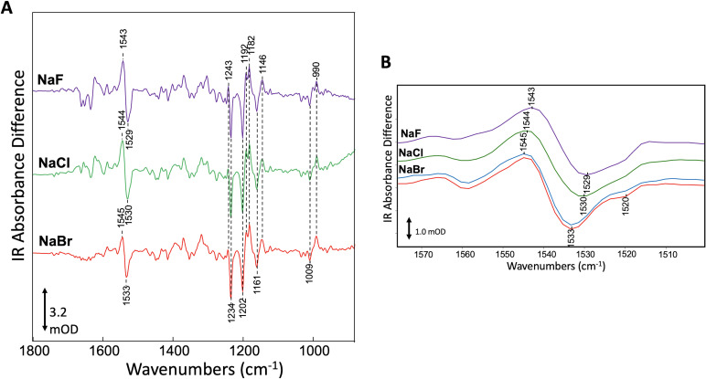
The crystal structure of the light-gated anion channel ACR1 reported in our previous Research Article (Li et al., 2019) revealed a continuous tunnel traversing the protein from extracellular to intracellular pores. We proposed the tunnel as the conductance channel closed by three constrictions: C1 in the extracellular half, mid-membrane C2 containing the photoactive site, and C3 on the cytoplasmic side. Reported here, the crystal structure of bromide-bound ACR1 reveals structural changes that relax the C1 and C3 constrictions, including a novel salt-bridge switch mechanism involving C1 and the photoactive site. These findings indicate that substrate binding induces a transition from an inactivated state to a pre-activated state in the dark that facilitates channel opening by reducing free energy in the tunnel constrictions. The results provide direct evidence that the tunnel is the closed form of the channel of ACR1 and shed light on the light-gated channel activation mechanism.

摘要

我们之前的研究文章(Li 等人,2019)中报道了光敏阴离子通道 ACR1 的晶体结构,该结构揭示了一条从细胞外到细胞内孔连续贯穿蛋白质的隧道。我们提出这条隧道是由三个限制因素关闭的电导通道:细胞外半部分的 C1、含有光活性位点的中膜 C2 和细胞质侧的 C3。本文报道了溴化物结合的 ACR1 的晶体结构,揭示了结构变化,这些变化放松了 C1 和 C3 的限制,包括涉及 C1 和光活性位点的新型盐桥开关机制。这些发现表明,底物结合诱导从无活性状态到预激活状态的转变,在黑暗中促进通道打开,减少隧道限制中的自由能。结果提供了直接证据表明隧道是 ACR1 通道的关闭形式,并阐明了光门控通道激活机制。

https://cdn.ncbi.nlm.nih.gov/pmc/blobs/1796/8172240/33fb03f956d9/elife-65903-fig4-figsupp1.jpg
https://cdn.ncbi.nlm.nih.gov/pmc/blobs/1796/8172240/5e18cf85ca94/elife-65903-fig1.jpg
https://cdn.ncbi.nlm.nih.gov/pmc/blobs/1796/8172240/ef52b7e89e22/elife-65903-fig1-figsupp1.jpg
https://cdn.ncbi.nlm.nih.gov/pmc/blobs/1796/8172240/7f03f2739831/elife-65903-fig2.jpg
https://cdn.ncbi.nlm.nih.gov/pmc/blobs/1796/8172240/c51c6883f124/elife-65903-fig3.jpg
https://cdn.ncbi.nlm.nih.gov/pmc/blobs/1796/8172240/99abc4448446/elife-65903-fig4.jpg
https://cdn.ncbi.nlm.nih.gov/pmc/blobs/1796/8172240/33fb03f956d9/elife-65903-fig4-figsupp1.jpg

相似文献

1
The crystal structure of bromide-bound ACR1 reveals a pre-activated state in the transmembrane anion tunnel.溴化物结合的 ACR1 晶体结构揭示了跨膜阴离子通道中的预激活状态。
Elife. 2021 May 17;10:e65903. doi: 10.7554/eLife.65903.
2
Crystal structure of a natural light-gated anion channelrhodopsin.天然光门控阴离子通道视紫红质的晶体结构。
Elife. 2019 Jan 7;8:e41741. doi: 10.7554/eLife.41741.
3
Crystal structure of the natural anion-conducting channelrhodopsin GtACR1.天然阴离子通道视紫红质 GtACR1 的晶体结构。
Nature. 2018 Sep;561(7723):343-348. doi: 10.1038/s41586-018-0511-6. Epub 2018 Aug 29.
4
Gating mechanisms of a natural anion channelrhodopsin.一种天然阴离子通道视紫红质的门控机制。
Proc Natl Acad Sci U S A. 2015 Nov 17;112(46):14236-41. doi: 10.1073/pnas.1513602112. Epub 2015 Nov 2.
5
Green-Sensitive, Long-Lived, Step-Functional Anion Channelrhodopsin-2 Variant as a High-Potential Neural Silencing Tool.对绿光敏感、长时程、阶跃功能的阴离子通道视紫红质-2 变体作为一种高潜力的神经沉默工具。
J Phys Chem Lett. 2020 Aug 6;11(15):6214-6218. doi: 10.1021/acs.jpclett.0c01406. Epub 2020 Jul 22.
6
Opposite Charge Movements Within the Photoactive Site Modulate Two-Step Channel Closing in GtACR1.光活性位点内的反荷移动调节 GtACR1 的两步通道关闭。
Biophys J. 2019 Nov 19;117(10):2034-2040. doi: 10.1016/j.bpj.2019.10.009. Epub 2019 Oct 14.
7
Structural mechanisms of selectivity and gating in anion channelrhodopsins.阴离子通道视紫红质的选择性和门控的结构机制。
Nature. 2018 Sep;561(7723):349-354. doi: 10.1038/s41586-018-0504-5. Epub 2018 Aug 29.
8
Exploring Protonation State, Ion Binding, and Photoactivated Channel Opening of an Anion Channelrhodopsin by Molecular Simulations.通过分子模拟探究阴离子通道视紫红质的质子化状态、离子结合和光激活通道开放。
J Phys Chem B. 2024 Sep 12;128(36):8613-8627. doi: 10.1021/acs.jpcb.4c03216. Epub 2024 Aug 29.
9
Proton transfer pathway in anion channelrhodopsin-1.阴离子通道视紫红质-1 中的质子转移途径。
Elife. 2021 Dec 21;10:e72264. doi: 10.7554/eLife.72264.
10
Time-resolved spectroscopic and electrophysiological data reveal insights in the gating mechanism of anion channelrhodopsin.时间分辨光谱和电生理数据揭示了阴离子通道视紫红质门控机制的见解。
Commun Biol. 2021 May 14;4(1):578. doi: 10.1038/s42003-021-02101-5.

引用本文的文献

1
Evolution of macromolecular crystallography beamlines at the Swiss Light Source and SwissFEL.瑞士光源和瑞士自由电子激光装置上大分子晶体学光束线的发展
J Synchrotron Radiat. 2025 Sep 1;32(Pt 5):1162-1183. doi: 10.1107/S1600577525005016. Epub 2025 Jul 14.
2
My remembrances of H.G. Khorana: exploring the mechanism of bacteriorhodopsin with site-directed mutagenesis and FTIR difference spectroscopy.我对H.G. 科拉纳的回忆:利用定点诱变和傅里叶变换红外差光谱法探索细菌视紫红质的机制。
Biophys Rev. 2023 Feb 6;15(1):103-110. doi: 10.1007/s12551-023-01046-9. eCollection 2023 Feb.
3
Structural Foundations of Potassium Selectivity in Channelrhodopsins.

本文引用的文献

1
Making a difference in multi-data-set crystallography: simple and deterministic data-scaling/selection methods.在多数据集晶体学中有所作为:简单且确定的数据缩放/选择方法。
Acta Crystallogr D Struct Biol. 2020 Jul 1;76(Pt 7):636-652. doi: 10.1107/S2059798320006348. Epub 2020 Jun 17.
2
Opposite Charge Movements Within the Photoactive Site Modulate Two-Step Channel Closing in GtACR1.光活性位点内的反荷移动调节 GtACR1 的两步通道关闭。
Biophys J. 2019 Nov 19;117(10):2034-2040. doi: 10.1016/j.bpj.2019.10.009. Epub 2019 Oct 14.
3
Automated data collection and real-time data analysis suite for serial synchrotron crystallography.
通道视紫红质中钾离子选择性的结构基础。
mBio. 2022 Dec 20;13(6):e0303922. doi: 10.1128/mbio.03039-22. Epub 2022 Nov 22.
4
Yeast as a tool for membrane protein production and structure determination.酵母作为生产膜蛋白和确定其结构的工具。
FEMS Yeast Res. 2022 Oct 20;22(1). doi: 10.1093/femsyr/foac047.
5
Rhodopsins: An Excitingly Versatile Protein Species for Research, Development and Creative Engineering.视紫红质:一种用于研究、开发和创新工程的极具通用性的蛋白质种类。
Front Chem. 2022 Jun 22;10:879609. doi: 10.3389/fchem.2022.879609. eCollection 2022.
6
Conformational alterations in unidirectional ion transport of a light-driven chloride pump revealed using X-ray free electron lasers.利用 X 射线自由电子激光揭示光驱动氯离子泵单向离子传输的构象变化。
Proc Natl Acad Sci U S A. 2022 Mar 1;119(9). doi: 10.1073/pnas.2117433119.
7
Emerging Diversity of Channelrhodopsins and Their Structure-Function Relationships.视紫红质通道蛋白的新多样性及其结构-功能关系。
Front Cell Neurosci. 2022 Jan 24;15:800313. doi: 10.3389/fncel.2021.800313. eCollection 2021.
8
Proton transfer pathway in anion channelrhodopsin-1.阴离子通道视紫红质-1 中的质子转移途径。
Elife. 2021 Dec 21;10:e72264. doi: 10.7554/eLife.72264.
9
Time-resolved spectroscopic and electrophysiological data reveal insights in the gating mechanism of anion channelrhodopsin.时间分辨光谱和电生理数据揭示了阴离子通道视紫红质门控机制的见解。
Commun Biol. 2021 May 14;4(1):578. doi: 10.1038/s42003-021-02101-5.
用于串行同步加速器晶体学的自动数据收集和实时数据分析套件。
J Synchrotron Radiat. 2019 Jan 1;26(Pt 1):244-252. doi: 10.1107/S1600577518016570.
4
Crystal structure of a natural light-gated anion channelrhodopsin.天然光门控阴离子通道视紫红质的晶体结构。
Elife. 2019 Jan 7;8:e41741. doi: 10.7554/eLife.41741.
5
In situ serial crystallography for rapid de novo membrane protein structure determination.用于快速从头测定膜蛋白结构的原位连续晶体学。
Commun Biol. 2018 Aug 27;1:124. doi: 10.1038/s42003-018-0123-6. eCollection 2018.
6
Crystal structure of the natural anion-conducting channelrhodopsin GtACR1.天然阴离子通道视紫红质 GtACR1 的晶体结构。
Nature. 2018 Sep;561(7723):343-348. doi: 10.1038/s41586-018-0511-6. Epub 2018 Aug 29.
7
DA+ data acquisition and analysis software at the Swiss Light Source macromolecular crystallography beamlines.瑞士光源大分子晶体学光束线处的DA+数据采集与分析软件。
J Synchrotron Radiat. 2018 Jan 1;25(Pt 1):293-303. doi: 10.1107/S1600577517014503.
8
Structural Changes in an Anion Channelrhodopsin: Formation of the K and L Intermediates at 80 K.一种阴离子通道视紫红质的结构变化:80K 时 K 和 L 中间体的形成
Biochemistry. 2017 Apr 25;56(16):2197-2208. doi: 10.1021/acs.biochem.7b00002. Epub 2017 Apr 10.
9
Crystal structure and functional characterization of a light-driven chloride pump having an NTQ motif.具有 NTQ 基序的光驱动氯离子泵的晶体结构和功能特征。
Nat Commun. 2016 Aug 24;7:12677. doi: 10.1038/ncomms12677.
10
Resonance Raman Study of an Anion Channelrhodopsin: Effects of Mutations near the Retinylidene Schiff Base.一种阴离子通道视紫红质的共振拉曼研究:视黄叉席夫碱附近突变的影响。
Biochemistry. 2016 Apr 26;55(16):2371-80. doi: 10.1021/acs.biochem.6b00104. Epub 2016 Apr 14.