Li Lei, Zhu Haogang, Zhang Zhenyu, Zhao Liang, Xu Liang, Jonas Rahul A, Garway-Heath David F, Jonas Jost B, Wang Ya Xing
State Key Laboratory of Software Development Environment, School of Computer Science and Engineering, Beihang University, Beijing, China.
Beijing Institute of Ophthalmology, Beijing Tongren Hospital, Capital University of Medical Science, Beijing Ophthalmology and Visual Sciences Key Laboratory, Beijing, China.
JMIR Med Inform. 2021 May 18;9(5):e22664. doi: 10.2196/22664.
Due to the axial elongation-associated changes in the optic nerve and retina in high myopia, traditional methods like optic disc evaluation and visual field are not able to correctly differentiate glaucomatous lesions. It has been clinically challenging to detect glaucoma in highly myopic eyes.
This study aimed to develop a neural network to adjust for the dependence of the peripapillary retinal nerve fiber layer (RNFL) thickness (RNFLT) profile on age, gender, and ocular biometric parameters and to evaluate the network's performance for glaucoma diagnosis, especially in high myopia.
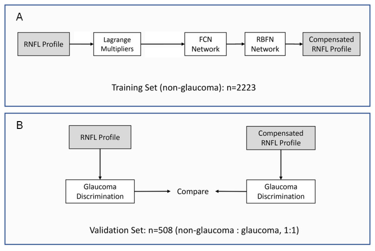
RNFLT with 768 points on the circumferential 3.4-mm scan was measured using spectral-domain optical coherence tomography. A fully connected network and a radial basis function network were trained for vertical (scaling) and horizontal (shift) transformation of the RNFLT profile with adjustment for age, axial length (AL), disc-fovea angle, and distance in a test group of 2223 nonglaucomatous eyes. The performance of RNFLT compensation was evaluated in an independent group of 254 glaucoma patients and 254 nonglaucomatous participants.
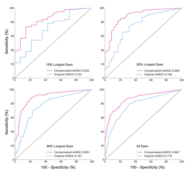
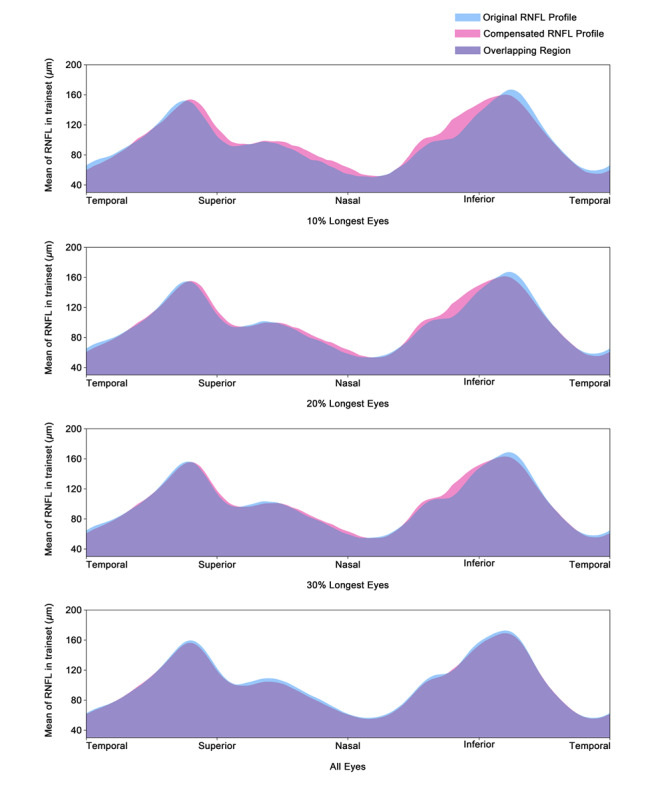
By applying the RNFL compensation algorithm, the area under the receiver operating characteristic curve for detecting glaucoma increased from 0.70 to 0.84, from 0.75 to 0.89, from 0.77 to 0.89, and from 0.78 to 0.87 for eyes in the highest 10% percentile subgroup of the AL distribution (mean 26.0, SD 0.9 mm), highest 20% percentile subgroup of the AL distribution (mean 25.3, SD 1.0 mm), highest 30% percentile subgroup of the AL distribution (mean 24.9, SD 1.0 mm), and any AL (mean 23.5, SD 1.2 mm), respectively, in comparison with unadjusted RNFLT. The difference between uncompensated and compensated RNFLT values increased with longer axial length, with enlargement of 19.8%, 18.9%, 16.2%, and 11.3% in the highest 10% percentile subgroup, highest 20% percentile subgroup, highest 30% percentile subgroup, and all eyes, respectively.
In a population-based study sample, an algorithm-based adjustment for age, gender, and ocular biometric parameters improved the diagnostic precision of the RNFLT profile for glaucoma detection particularly in myopic and highly myopic eyes.
由于高度近视患者视神经和视网膜存在与眼轴延长相关的变化,传统的视盘评估和视野检查等方法无法准确区分青光眼性病变。在高度近视眼中检测青光眼一直是临床难题。
本研究旨在开发一种神经网络,以调整视乳头周围视网膜神经纤维层(RNFL)厚度(RNFLT)分布与年龄、性别及眼部生物测量参数的相关性,并评估该网络在青光眼诊断中的性能,尤其是在高度近视眼中的性能。
使用光谱域光学相干断层扫描测量3.4毫米圆周扫描上768个点的RNFLT。在一个包含2223只非青光眼性眼的测试组中,训练一个全连接网络和一个径向基函数网络,用于对RNFLT分布进行垂直(缩放)和水平(移位)变换,并根据年龄、眼轴长度(AL)、视盘-黄斑中心凹夹角和距离进行调整。在一个独立的包含254例青光眼患者和254例非青光眼参与者的组中评估RNFLT补偿的性能。
应用RNFL补偿算法后,在AL分布最高的10%百分位子组(平均26.0,标准差0.9毫米)、AL分布最高的20%百分位子组(平均25.3,标准差1.0毫米)、AL分布最高的30%百分位子组(平均24.9,标准差1.0毫米)以及任何AL(平均23.5,标准差1.2毫米)的眼中,检测青光眼的受试者工作特征曲线下面积分别从未调整的RNFLT时的0.70增加到0.84、从0.75增加到0.89、从0.77增加到0.89以及从0.78增加到0.87。未补偿和补偿后的RNFLT值之间的差异随眼轴长度增加而增大,在最高的10%百分位子组、最高的20%百分位子组、最高的30%百分位子组以及所有眼中分别增大19.8%、18.9%、16.2%和11.3%。
在一项基于人群的研究样本中,基于算法对年龄、性别和眼部生物测量参数进行调整可提高RNFLT分布在青光眼检测中的诊断精度,尤其是在近视和高度近视眼中。