Leibniz Research Centre for Working Environment and Human Factors, Technical University Dortmund, Ardeystr. 67, 44139, Dortmund, Germany.
Department of Forensic Medicine and Toxicology, Faculty of Veterinary Medicine, South Valley University, Qena, 83523, Egypt.
Arch Toxicol. 2021 Jun;95(6):2163-2177. doi: 10.1007/s00204-021-03073-5. Epub 2021 May 18.
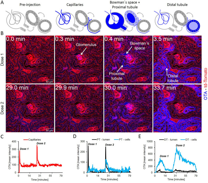
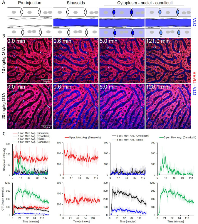
Local accumulation of xenobiotics in human and animal tissues may cause adverse effects. Large differences in their concentrations may exist between individual cell types, often due to the expression of specific uptake and export carriers. Here we established a two-photon microscopy-based technique for spatio-temporal detection of the distribution of mycotoxins in intact kidneys and livers of anesthetized mice with subcellular resolution. The mycotoxins ochratoxin A (OTA, 10 mg/kg b.w.) and aflatoxin B (AFB, 1.5 mg/kg b.w.), which both show blue auto-fluorescence, were analyzed after intravenous bolus injections. Within seconds after administration, OTA was filtered by glomeruli, and enriched in distal tubular epithelial cells (dTEC). A striking feature of AFB toxicokinetics was its very rapid uptake from sinusoidal blood into hepatocytes (t ~ 4 min) and excretion into bile canaliculi. Interestingly, AFB was enriched in the nuclei of hepatocytes with zonal differences in clearance. In the cytoplasm of pericentral hepatocytes, the half-life (t~ 63 min) was much longer compared to periportal hepatocytes of the same lobules (t ~ 9 min). In addition, nuclear AFB from periportal hepatocytes cleared faster compared to the pericentral region. These local differences in AFB clearance may be due to the pericentral expression of cytochrome P450 enzymes that activate AFB to protein- and DNA-binding metabolites. In conclusion, the present study shows that large spatio-temporal concentration differences exist within the same tissues and its analysis may provide valuable additional information to conventional toxicokinetic studies.
外源化学物在人体和动物组织中的局部积累可能会导致不良反应。由于特定摄取和外排载体的表达,不同细胞类型之间的浓度可能存在很大差异。在这里,我们建立了一种基于双光子显微镜的技术,用于以亚细胞分辨率时空检测麻醉小鼠完整肾脏和肝脏中真菌毒素的分布。分析了静脉内推注后具有蓝色自发荧光的两种真菌毒素黄曲霉毒素 A(OTA,10mg/kg b.w.)和黄曲霉毒素 B(AFB,1.5mg/kg b.w.)。给药后几秒钟内,OTA 被肾小球滤过,并在远端管状上皮细胞(dTEC)中富集。AFB 毒代动力学的一个显著特征是其从窦状血液中非常快速地摄取并排入胆小管。有趣的是,AFB 在肝细胞的细胞核中富集,清除存在区域差异。在中央周围肝细胞的细胞质中,半衰期(t63 分钟)比同一小叶的门周肝细胞(t9 分钟)长得多。此外,与中央区相比,门周肝细胞的核 AFB 清除速度更快。这些 AFB 清除的局部差异可能是由于细胞色素 P450 酶在中央周围表达,将 AFB 激活为蛋白和 DNA 结合代谢物。总之,本研究表明,同一组织内存在很大的时空浓度差异,其分析可能为传统毒代动力学研究提供有价值的附加信息。