• 文献检索
  • 文档翻译
  • 深度研究
  • 学术资讯
  • Suppr Zotero 插件Zotero 插件
  • 邀请有礼
  • 套餐&价格
  • 历史记录
应用&插件
Suppr Zotero 插件Zotero 插件浏览器插件Mac 客户端Windows 客户端微信小程序
定价
高级版会员购买积分包购买API积分包
服务
文献检索文档翻译深度研究API 文档MCP 服务
关于我们
关于 Suppr公司介绍联系我们用户协议隐私条款
关注我们

Suppr 超能文献

核心技术专利:CN118964589B侵权必究
粤ICP备2023148730 号-1Suppr @ 2026

文献检索

告别复杂PubMed语法,用中文像聊天一样搜索,搜遍4000万医学文献。AI智能推荐,让科研检索更轻松。

立即免费搜索

文件翻译

保留排版,准确专业,支持PDF/Word/PPT等文件格式,支持 12+语言互译。

免费翻译文档

深度研究

AI帮你快速写综述,25分钟生成高质量综述,智能提取关键信息,辅助科研写作。

立即免费体验

多发性硬化症患者的角膜免疫细胞增加。

Corneal Immune Cells Are Increased in Patients With Multiple Sclerosis.

机构信息

Weill Cornell Medicine-Qatar, Research Division, Doha, Qatar.

Department of Neurology, University of Pittsburgh Medical Center, Pittsburgh, PA, USA.

出版信息

Transl Vis Sci Technol. 2021 Apr 1;10(4):19. doi: 10.1167/tvst.10.4.19.

DOI:10.1167/tvst.10.4.19
PMID:34003997
原文链接:https://pmc.ncbi.nlm.nih.gov/articles/PMC8083118/
Abstract

PURPOSE

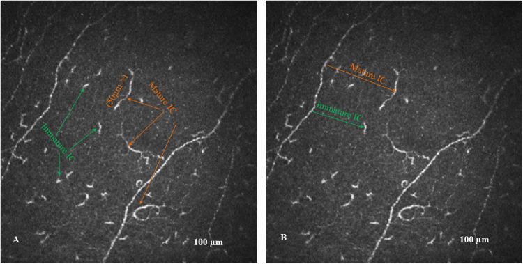
Corneal confocal microscopy (CCM) is an ophthalmic imaging technique that has been used to identify increased corneal immune cells in patients with immune-mediated peripheral neuropathy. Given that multiple sclerosis has an immune-mediated etiology, we have compared corneal immune cell (IC) density and near-nerve distance in different subtypes of patients with multiple sclerosis (MS) to controls.

METHODS

This is a blinded, cross-sectional study conducted at a tertiary hospital. Patients with clinically isolated syndrome (CIS) (n = 9), relapsing-remitting multiple sclerosis (RRMS) (n = 43), secondary progressive multiple sclerosis (SPMS) (n = 22), and control subjects (n = 20) underwent CCM. The total, mature, and immature corneal IC density and their nearest nerve distance were quantified.

RESULTS

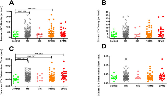
The total IC density was higher in patients with MS (P = 0.02), RRMS (P = 0.01), and SPMS (P = 0.04) but not CIS (P = 0.99) compared to controls. Immature IC density was higher in patients with MS (P = 0.03) and RRMS (P = 0.02) but not SPMS (P = 0.10) or CIS (P = 0.99) compared to controls. Mature IC density (P = 0.15) did not differ between patients with MS and controls. The immature IC near-nerve distance was significantly greater in patients with MS (P = 0.001), RRMS (P = 0.007), and SPMS (P = 0.002) compared to controls. Immature IC density correlated with the Symbol Digit Modalities Test (r = -0.281, P = 0.02) and near-nerve distance correlated with the Expanded Disability Status Scale (r = 0.289, P = 0.005).

CONCLUSIONS

In vivo CCM demonstrates an increase in immature IC density and the near-nerve distance in patients with MS. These observations merit further studies to assess the utility of CCM in assessing neuroimmune alterations in MS.

TRANSLATIONAL RELEVANCE

Multiple sclerosis is an immune-mediated neurodegenerative disease. Dendritic cells mediate communication between the innate and adaptive immune systems. We have used in vivo CCM to show increased corneal ICs and suggest it may act as an imaging biomarker for disease status in patients with MS.

摘要

目的

角膜共聚焦显微镜(CCM)是一种眼科成像技术,已用于识别免疫介导的周围神经病患者中角膜免疫细胞的增加。鉴于多发性硬化症具有免疫介导的病因,我们比较了不同亚型多发性硬化症(MS)患者与对照组之间的角膜免疫细胞(IC)密度和近神经距离。

方法

这是在一家三级医院进行的一项盲法、横断面研究。临床孤立综合征(CIS)患者(n=9)、复发缓解型多发性硬化症(RRMS)患者(n=43)、继发进展型多发性硬化症(SPMS)患者(n=22)和对照组(n=20)接受了 CCM。定量分析总、成熟和不成熟角膜 IC 密度及其最近神经距离。

结果

与对照组相比,MS(P=0.02)、RRMS(P=0.01)和 SPMS(P=0.04)患者的总 IC 密度更高,但 CIS 患者(P=0.99)则不然。MS(P=0.03)和 RRMS(P=0.02)患者的不成熟 IC 密度更高,但 SPMS(P=0.10)或 CIS(P=0.99)患者则不然。MS 患者与对照组之间的成熟 IC 密度无差异(P=0.15)。MS(P=0.001)、RRMS(P=0.007)和 SPMS(P=0.002)患者的不成熟 IC 近神经距离明显大于对照组。不成熟 IC 密度与符号数字模态测试(r=-0.281,P=0.02)相关,近神经距离与扩展残疾状况量表(r=0.289,P=0.005)相关。

结论

体内 CCM 显示 MS 患者不成熟 IC 密度和近神经距离增加。这些观察结果值得进一步研究,以评估 CCM 在评估 MS 神经免疫改变中的效用。

翻译的准确性和流畅度如何?如果有需要改进的地方,请提供具体建议。

https://cdn.ncbi.nlm.nih.gov/pmc/blobs/f445/8083118/57481d939e97/tvst-10-4-19-f002.jpg
https://cdn.ncbi.nlm.nih.gov/pmc/blobs/f445/8083118/decc70439fd5/tvst-10-4-19-f001.jpg
https://cdn.ncbi.nlm.nih.gov/pmc/blobs/f445/8083118/57481d939e97/tvst-10-4-19-f002.jpg
https://cdn.ncbi.nlm.nih.gov/pmc/blobs/f445/8083118/decc70439fd5/tvst-10-4-19-f001.jpg
https://cdn.ncbi.nlm.nih.gov/pmc/blobs/f445/8083118/57481d939e97/tvst-10-4-19-f002.jpg

相似文献

1
Corneal Immune Cells Are Increased in Patients With Multiple Sclerosis.多发性硬化症患者的角膜免疫细胞增加。
Transl Vis Sci Technol. 2021 Apr 1;10(4):19. doi: 10.1167/tvst.10.4.19.
2
Use of Corneal Confocal Microscopy to Detect Corneal Nerve Loss and Increased Dendritic Cells in Patients With Multiple Sclerosis.使用角膜共焦显微镜检测多发性硬化症患者的角膜神经损伤和树突状细胞增多
JAMA Ophthalmol. 2017 Jul 1;135(7):777-782. doi: 10.1001/jamaophthalmol.2017.1590.
3
Corneal immune cells as a biomarker of inflammation in multiple sclerosis: a longitudinal study.角膜免疫细胞作为多发性硬化症炎症的生物标志物:一项纵向研究。
Ther Adv Neurol Disord. 2023 Oct 29;16:17562864231204974. doi: 10.1177/17562864231204974. eCollection 2023.
4
Corneal axonal loss as an imaging biomarker of neurodegeneration in multiple sclerosis: a longitudinal study.角膜轴突丢失作为多发性硬化症神经退行性变的影像学生物标志物:一项纵向研究。
Ther Adv Neurol Disord. 2023 Feb 6;16:17562864221118731. doi: 10.1177/17562864221118731. eCollection 2023.
5
IL17 and IFNgamma production by peripheral blood mononuclear cells from clinically isolated syndrome to secondary progressive multiple sclerosis.从临床孤立综合征到继发进展型多发性硬化症患者外周血单个核细胞产生白细胞介素17和γ干扰素的情况
Cytokine. 2008 Oct;44(1):22-5. doi: 10.1016/j.cyto.2008.08.007. Epub 2008 Sep 14.
6
Corneal Confocal Microscopy: An Imaging Endpoint for Axonal Degeneration in Multiple Sclerosis.角膜共焦显微镜检查:多发性硬化症轴突退变的一种成像终点指标
Invest Ophthalmol Vis Sci. 2017 Jul 1;58(9):3677-3681. doi: 10.1167/iovs.17-22050.
7
Clinically relevant cranio-caudal patterns of cervical cord atrophy evolution in MS.MS 中颈髓萎缩演变的临床相关头尾模式。
Neurology. 2019 Nov 12;93(20):e1852-e1866. doi: 10.1212/WNL.0000000000008466. Epub 2019 Oct 14.
8
[Information processing speed and influential factors in multiple sclerosis].[多发性硬化症中的信息处理速度及影响因素]
Zhonghua Yi Xue Za Zhi. 2016 Apr 19;96(15):1173-7. doi: 10.3760/cma.j.issn.0376-2491.2016.15.006.
9
Adhesion molecules in multiple sclerosis: relation to subtypes of disease and methylprednisolone therapy.多发性硬化症中的黏附分子:与疾病亚型及甲基强的松龙治疗的关系
Arch Neurol. 2000 Apr;57(4):546-51. doi: 10.1001/archneur.57.4.546.
10
Secondary progressive multiple sclerosis: a systematic review of costs and health state utilities.继发进展型多发性硬化症:成本和健康状态效用的系统评价。
Curr Med Res Opin. 2021 Jun;37(6):995-1004. doi: 10.1080/03007995.2021.1904860. Epub 2021 Apr 6.

引用本文的文献

1
Oculomics analysis in multiple sclerosis: Current ophthalmic clinical and imaging biomarkers.眼科学分析在多发性硬化症中的应用:当前的眼科临床和影像学生物标志物。
Eye (Lond). 2024 Oct;38(14):2701-2710. doi: 10.1038/s41433-024-03132-y. Epub 2024 Jun 10.
2
Neuroimaging to monitor worsening of multiple sclerosis: advances supported by the grant for multiple sclerosis innovation.用于监测多发性硬化症病情恶化的神经影像学:多发性硬化症创新基金支持下的进展
Front Neurol. 2023 Dec 1;14:1319869. doi: 10.3389/fneur.2023.1319869. eCollection 2023.
3
Corneal immune cells as a biomarker of inflammation in multiple sclerosis: a longitudinal study.

本文引用的文献

1
Mechanisms underlying progression in multiple sclerosis.多发性硬化症进展的机制。
Curr Opin Neurol. 2020 Jun;33(3):277-285. doi: 10.1097/WCO.0000000000000818.
2
Reduced association between dendritic cells and corneal sub-basal nerve fibers in patients with fibromyalgia syndrome.纤维肌痛综合征患者树突状细胞与角膜基底神经纤维的关联减少。
J Peripher Nerv Syst. 2020 Mar;25(1):9-18. doi: 10.1111/jns.12360. Epub 2020 Jan 2.
3
Early corneal nerve fibre damage and increased Langerhans cell density in children with type 1 diabetes mellitus.
角膜免疫细胞作为多发性硬化症炎症的生物标志物:一项纵向研究。
Ther Adv Neurol Disord. 2023 Oct 29;16:17562864231204974. doi: 10.1177/17562864231204974. eCollection 2023.
4
In-vivo corneal confocal microscopy: Imaging analysis, biological insights and future directions.体内角膜共聚焦显微镜:成像分析、生物学见解和未来方向。
Commun Biol. 2023 Jun 19;6(1):652. doi: 10.1038/s42003-023-05005-8.
5
Corneal nerve loss and increased Langerhans cells are associated with disease severity in patients with rheumatoid arthritis.角膜神经丢失和朗格汉斯细胞增多与类风湿关节炎患者的疾病严重程度相关。
Eye (Lond). 2023 Oct;37(14):2950-2955. doi: 10.1038/s41433-023-02447-6. Epub 2023 Feb 18.
6
Diurnal changes and topographical distribution of ocular surface epithelial dendritic cells in humans, and repeatability of density and morphology assessment.人类眼表上皮树突状细胞的昼夜变化和分布特征,以及密度和形态评估的可重复性。
Ophthalmic Physiol Opt. 2023 Mar;43(2):273-283. doi: 10.1111/opo.13087. Epub 2023 Jan 2.
7
In vivo corneal confocal microscopy as a non-invasive test to assess obesity induced small fibre nerve damage and inflammation.体内角膜共聚焦显微镜作为一种非侵入性测试,可评估肥胖引起的小纤维神经损伤和炎症。
Eye (Lond). 2023 Aug;37(11):2226-2232. doi: 10.1038/s41433-022-02321-x. Epub 2022 Nov 28.
8
Corneal Langerhans cells in children with celiac disease.儿童乳糜泻患者的角膜朗格汉斯细胞。
Sci Rep. 2022 Oct 31;12(1):18289. doi: 10.1038/s41598-022-22376-w.
9
Segmentation and Evaluation of Corneal Nerves and Dendritic Cells From In Vivo Confocal Microscopy Images Using Deep Learning.基于深度学习的活体共聚焦显微镜图像中角膜神经和树突状细胞的分割和评估。
Transl Vis Sci Technol. 2022 Jun 1;11(6):24. doi: 10.1167/tvst.11.6.24.
10
Defining an Optimal Sample Size for Corneal Epithelial Immune Cell Analysis Using Confocal Microscopy Images.利用共聚焦显微镜图像确定角膜上皮免疫细胞分析的最佳样本量
Front Med (Lausanne). 2022 Jun 1;9:848776. doi: 10.3389/fmed.2022.848776. eCollection 2022.
儿童 1 型糖尿病患者早期角膜神经纤维损伤和朗格汉斯细胞密度增加。
Sci Rep. 2019 Jun 19;9(1):8758. doi: 10.1038/s41598-019-45116-z.
4
Corneal confocal microscopy detects corneal nerve damage and increased dendritic cells in Fabry disease.角膜共焦显微镜检测法可发现法布里病的角膜神经损伤和树突状细胞增多。
Sci Rep. 2018 Aug 16;8(1):12244. doi: 10.1038/s41598-018-30688-z.
5
Confocal Microscopic Evaluation of Corneal Nerve Fibers and Dendritic Cells in Patients With Behçet's Disease.白塞病患者角膜神经纤维和树突状细胞的共聚焦显微镜评估
Front Neurol. 2018 Mar 28;9:204. doi: 10.3389/fneur.2018.00204. eCollection 2018.
6
Diagnosis of multiple sclerosis: 2017 revisions of the McDonald criteria.多发性硬化症的诊断:2017 年麦当劳标准修订版。
Lancet Neurol. 2018 Feb;17(2):162-173. doi: 10.1016/S1474-4422(17)30470-2. Epub 2017 Dec 21.
7
CIDP and other inflammatory neuropathies in diabetes - diagnosis and management.糖尿病相关 CIDP 和其他炎性神经病——诊断与管理。
Nat Rev Neurol. 2017 Oct;13(10):599-611. doi: 10.1038/nrneurol.2017.123. Epub 2017 Sep 15.
8
Corneal Confocal Microscopy: An Imaging Endpoint for Axonal Degeneration in Multiple Sclerosis.角膜共焦显微镜检查:多发性硬化症轴突退变的一种成像终点指标
Invest Ophthalmol Vis Sci. 2017 Jul 1;58(9):3677-3681. doi: 10.1167/iovs.17-22050.
9
Use of Corneal Confocal Microscopy to Detect Corneal Nerve Loss and Increased Dendritic Cells in Patients With Multiple Sclerosis.使用角膜共焦显微镜检测多发性硬化症患者的角膜神经损伤和树突状细胞增多
JAMA Ophthalmol. 2017 Jul 1;135(7):777-782. doi: 10.1001/jamaophthalmol.2017.1590.
10
Validity of the Symbol Digit Modalities Test as a cognition performance outcome measure for multiple sclerosis.符号数字模态测验作为多发性硬化症认知功能结果测量指标的效度
Mult Scler. 2017 Apr;23(5):721-733. doi: 10.1177/1352458517690821. Epub 2017 Feb 16.