Institute for Regenerative Medicine, Shanghai East Hospital, School of Life Sciences and Technology, Tongji University, Shanghai, People's Republic of China.
Research Center for Translational Medicine, Shanghai East Hospital, School of Medicine, Tongji University, Shanghai, People's Republic of China.
Stem Cell Res Ther. 2021 May 20;12(1):295. doi: 10.1186/s13287-021-02366-x.
Mesenchymal stem cells (MSCs) can improve cutaneous wound healing via the secretion of growth factors. However, the therapeutic efficacy of MSCs varies depending upon their source. Induced pluripotent stem cells are emerging as a promising source of MSCs with the potential to overcome several limitations of adult MSCs. This study compared the effectiveness of conditioned medium of MSCs derived from induced pluripotent stem cells (iMSC-CdM) with that derived from umbilical cord MSCs (uMSC-CdM) in a mouse cutaneous wound healing model. We also investigated the mechanisms of protection.
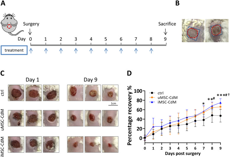
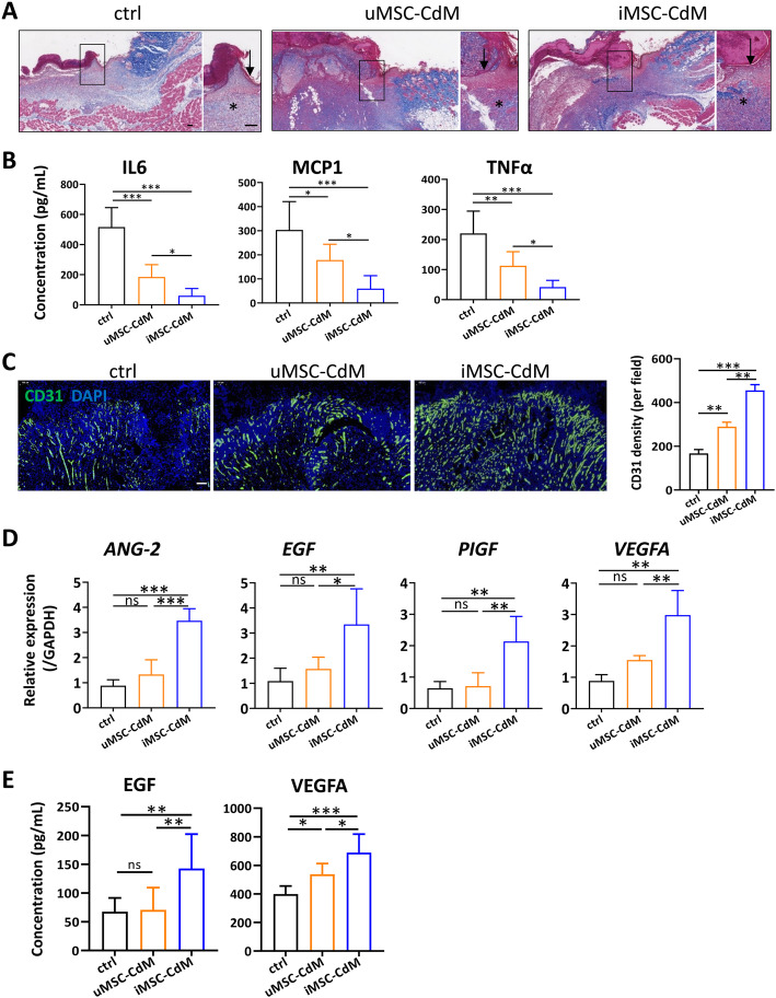
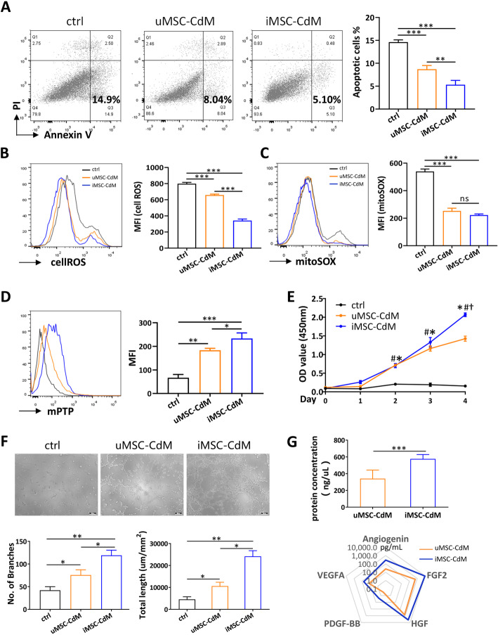
The iMSC-CdM or uMSC-CdM were topically applied to mice cutaneous wound model. The recovery rate, scar formation, inflammation and angiogenesis were measured. We compared angiogenesis cytokine expression between iMSC-CdM and uMSC-CdM and their protective effects on human umbilical vein endothelial cells (HUVECs) under HO-induced injury. The effects of iMSC-CdM on energy metabolism, mitochondria fragmentation and apoptosis were measured.
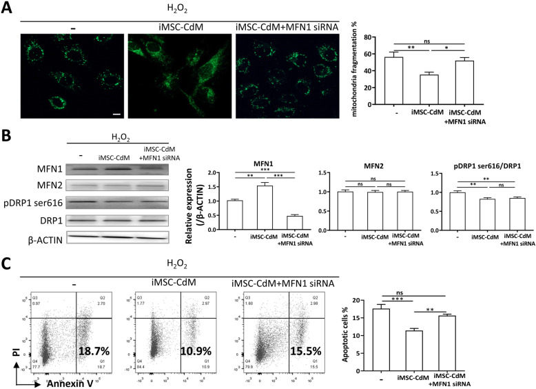
Topical application of iMSC-CdM was superior to the uMSC-CdM in accelerating wound closure and enhancing angiogenesis. Expression levels of angiogenetic cytokines were higher in iMSC-CdM than they were in uMSC-CdM. The iMSC-CdM protected HUVECs from HO induced injury more effectively than uMSC-CdM did. Administration of iMSC-CdM stimulated HUVEC proliferation, tube formation and energy metabolism via the ERK pathway. Mechanistically, iMSC-CdM inhibited HO-induced mitochondrial fragmentation and apoptosis of HUVECs.
Collectively, these findings indicate that iMSC-CdM is more effective than uMSC-CdM in treating cutaneous wounds, and in this way, iMSC-CdM may serve as a more constant and sustainable source for cell-free therapeutic approach.
间充质干细胞(MSCs)可通过分泌生长因子来改善皮肤伤口愈合。然而,MSCs 的治疗效果因来源而异。诱导多能干细胞作为 MSCs 的一种有前途的来源正在出现,具有克服成体 MSCs 若干局限性的潜力。本研究比较了诱导多能干细胞来源的条件培养基(iMSC-CdM)与脐带间充质干细胞来源的条件培养基(uMSC-CdM)在小鼠皮肤伤口愈合模型中的疗效。我们还研究了保护机制。
将 iMSC-CdM 或 uMSC-CdM 局部应用于小鼠皮肤伤口模型。测量恢复率、瘢痕形成、炎症和血管生成。我们比较了 iMSC-CdM 与 uMSC-CdM 之间的血管生成细胞因子表达及其在 HO 诱导损伤下对人脐静脉内皮细胞(HUVECs)的保护作用。测量 iMSC-CdM 对能量代谢、线粒体碎片化和细胞凋亡的影响。
iMSC-CdM 局部应用在加速伤口闭合和增强血管生成方面优于 uMSC-CdM。iMSC-CdM 的血管生成细胞因子表达水平高于 uMSC-CdM。iMSC-CdM 比 uMSC-CdM 更有效地保护 HUVECs 免受 HO 诱导的损伤。iMSC-CdM 通过 ERK 通路刺激 HUVEC 增殖、管形成和能量代谢。在机制上,iMSC-CdM 抑制 HO 诱导的 HUVEC 线粒体碎片化和凋亡。
总之,这些发现表明 iMSC-CdM 在治疗皮肤伤口方面比 uMSC-CdM 更有效,并且 iMSC-CdM 可能成为更恒定和可持续的无细胞治疗方法的来源。