Institute for Advanced Research (IAR), Nagoya University, Furo-cho, Chikusa-ku, Nagoya, Aichi 464-8601, Japan.
Institute of Transformative Bio-Molecules (WPI-ITbM), Nagoya University, Furo-cho, Chikusa-ku, Nagoya, Aichi 464-8601, Japan.
Plant Cell Physiol. 2021 Nov 10;62(8):1224-1230. doi: 10.1093/pcp/pcab062.
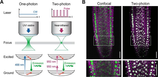
Live and deep imaging play a significant role in the physiological and biological study of organisms. Two-photon excitation microscopy (2PEM), also known as multiphoton excitation microscopy, is a fluorescent imaging technique that allows deep imaging of living tissues. Two-photon lasers use near-infrared (NIR) pulse lasers that are less invasive and permit deep tissue penetration. In this review, recent advances in two-photon imaging and their applications in plant studies are discussed. Compared to confocal microscopy, NIR 2PEM exhibits reduced plant-specific autofluorescence, thereby achieving greater depth and high-resolution imaging in plant tissues. Fluorescent proteins with long emission wavelengths, such as orange-red fluorescent proteins, are particularly suitable for two-photon live imaging in plants. Furthermore, deep- and high-resolution imaging was achieved using plant-specific clearing methods. In addition to imaging, optical cell manipulations can be performed using femtosecond pulsed lasers at the single cell or organelle level. Optical surgery and manipulation can reveal cellular communication during development. Advances in in vivo imaging using 2PEM will greatly benefit biological studies in plant sciences.
活体和深层成像在生物体的生理和生物学研究中发挥着重要作用。双光子激发显微镜(2PEM),也称为多光子激发显微镜,是一种荧光成像技术,可实现对活体组织的深层成像。双光子激光器使用近红外(NIR)脉冲激光器,其侵入性较小,允许深层组织穿透。本文综述了双光子成像的最新进展及其在植物研究中的应用。与共聚焦显微镜相比,NIR 2PEM 表现出较低的植物特异性自发荧光,从而在植物组织中实现更大的深度和高分辨率成像。发射波长较长的荧光蛋白,如橙红色荧光蛋白,特别适合植物的双光子活体成像。此外,使用植物特异性透明化方法实现了深层和高分辨率成像。除成像外,还可以在单细胞或细胞器水平使用飞秒脉冲激光进行光学细胞操作。光学手术和操作可以揭示发育过程中的细胞通讯。使用 2PEM 进行体内成像的进展将极大地有益于植物科学中的生物学研究。