Gangadaran Prakash, Li Xiu Juan, Lee Ho Won, Oh Ji Min, Kalimuthu Senthilkumar, Rajendran Ramya Lakshmi, Son Seung Hyun, Baek Se Hwan, Singh Thoudam Debraj, Zhu Liya, Jeong Shin Young, Lee Sang-Woo, Lee Jaetae, Ahn Byeong-Cheol
Department of Nuclear Medicine, Kyungpook National University School of Medicine and Hospital, Daegu 700-721, Republic of Korea.
Department of Medical Oncology, All India Institute of Medical Sciences (AIIMS), Ansari Nagar, New Delhi 110029, India.
Oncotarget. 2017 Nov 18;8(66):109894-109914. doi: 10.18632/oncotarget.22493. eCollection 2017 Dec 15.
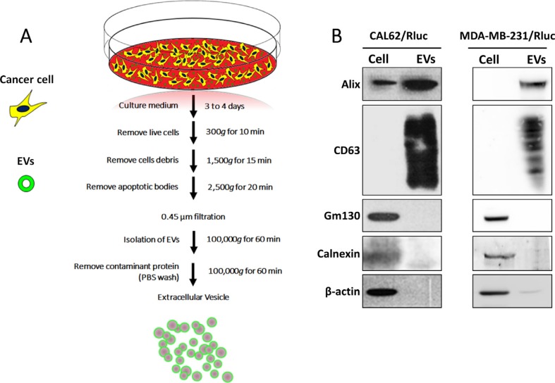
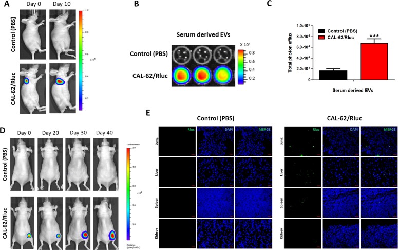
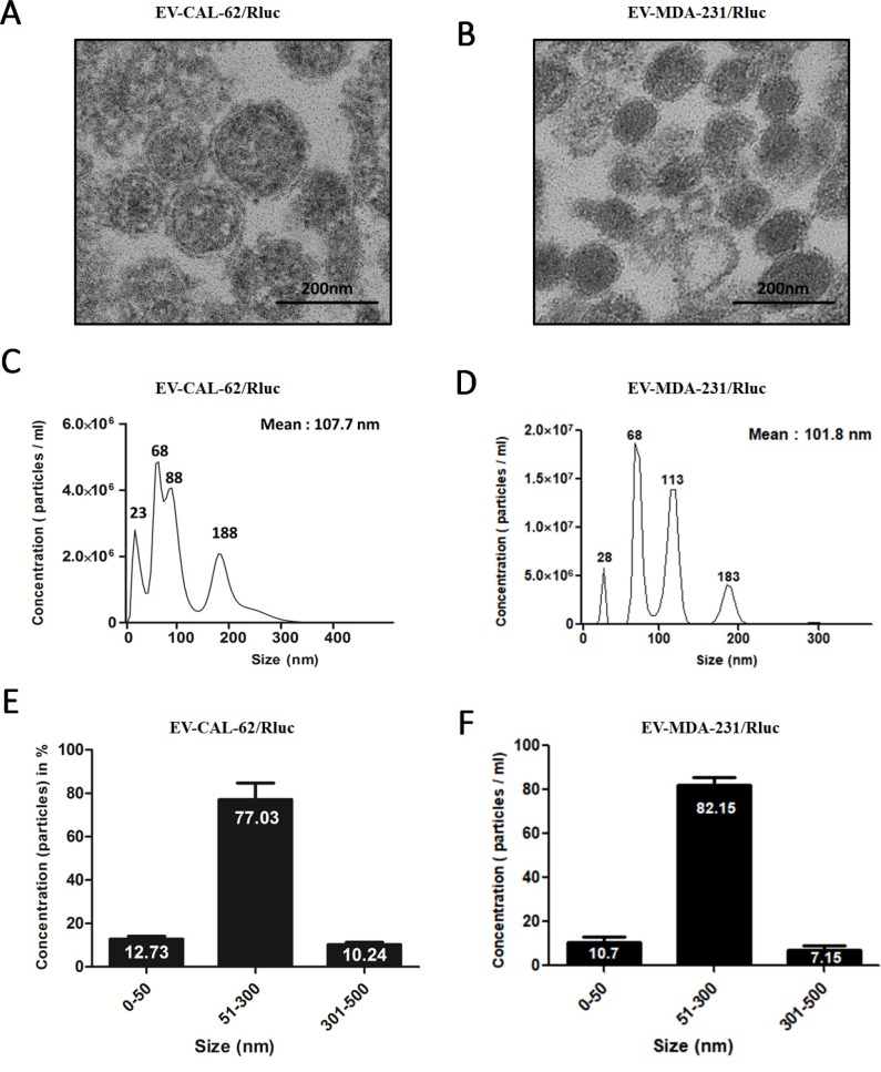
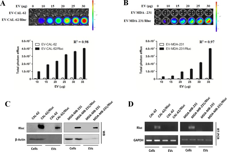
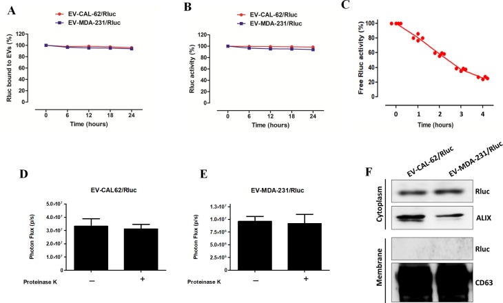
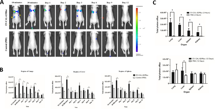
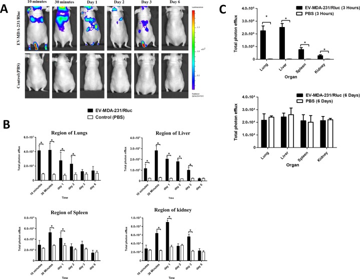
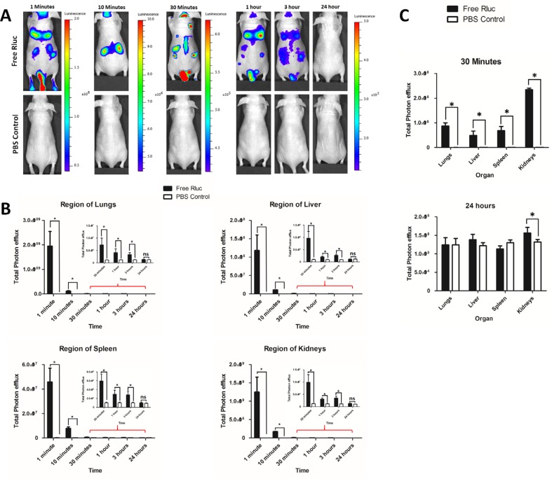
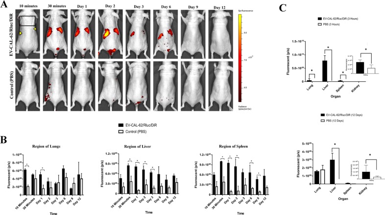
biodistribution and fate of extracellular vesicles (EVs) are still largely unknown and require reliable tracking techniques. In this study, bioluminescence imaging (BLI) using Renilla luciferase (Rluc) was developed and applied to monitoring of EVs derived from thyroid cancer (CAL-62 cells) and breast cancer (MDA-MB-231) in nude mice after intravenous administration and was compared with a dye-based labeling method for EV derived from CAL-62 cells. The EVs were successfully labeled with Rluc and visualized by BLI in mice. distribution of the EVs, as measured by BLI, was consistent with the results of organ analysis. EV-CAL-62/Rluc showed strong signals at lung followed by liver, spleen & kidney ( < 0.05). EV-MDA-MB-231/Rluc showed strong signals at liver followed by lung, spleen & kidney ( < 0.05). EV-CAL-62/Rluc and EV-MDA-MB-231/Rluc stayed in animal till day 9 and 3, respectively; showed a differential distribution. Spontaneous EV-CAL-62/Rluc shown distributed mostly to lung followed by liver, spleen & kidney. The new BLI system used to show spontaneous distribution of EV-CAL-62/Rluc in subcutaneous CAL-62/Rluc bearing mice. Dye (DiR)-labeled EV-CAL-62/Rluc showed a different distribution & compared to EV-CAL-62/Rluc. Fluorescent signals were predominately detected in the liver ( < 0.05) and spleen ( < 0.05) regions. The bioluminescent EVs developed in this study may be used for monitoring of EVs . This novel reporter-imaging approach to visualization of EVs in real time is expected to pave the way for monitoring of EVs in EV-based treatments.
细胞外囊泡(EVs)的生物分布和归宿在很大程度上仍不清楚,需要可靠的追踪技术。在本研究中,开发了使用海肾荧光素酶(Rluc)的生物发光成像(BLI),并将其应用于监测静脉注射后裸鼠体内源自甲状腺癌(CAL-62细胞)和乳腺癌(MDA-MB-231)的EVs,并与源自CAL-62细胞的EVs的基于染料的标记方法进行比较。EVs成功用Rluc标记,并通过BLI在小鼠体内可视化。通过BLI测量的EVs分布与器官分析结果一致。EV-CAL-62/Rluc在肺部显示出强烈信号,其次是肝脏、脾脏和肾脏(<0.05)。EV-MDA-MB-231/Rluc在肝脏显示出强烈信号,其次是肺部、脾脏和肾脏(<0.05)。EV-CAL-62/Rluc和EV-MDA-MB-231/Rluc分别在动物体内停留至第9天和第3天;显示出不同的分布。自发的EV-CAL-62/Rluc大多分布到肺部,其次是肝脏、脾脏和肾脏。新的BLI系统用于显示皮下携带CAL-62/Rluc的小鼠中EV-CAL-62/Rluc的自发分布。染料(DiR)标记的EV-CAL-62/Rluc显示出与EV-CAL-62/Rluc不同的分布。荧光信号主要在肝脏(<0.05)和脾脏(<0.05)区域检测到。本研究中开发的生物发光EVs可用于监测EVs。这种用于实时可视化EVs的新型报告基因成像方法有望为基于EVs的治疗中监测EVs铺平道路。