Emamian Farnoosh, Mahdipour Mostafa, Noori Khadijeh, Rostampour Masoumeh, Mousavi S Bentolhoda, Khazaie Habibolah, Khodaie-Ardakani Mohammadreza, Tahmasian Masoud, Zarei Mojtaba
Department of Psychiatry, University of Social Welfare and Rehabilitation Sciences, Tehran, Iran.
Sleep Disorders Research Center, Kermanshah University of Medical Sciences, Kermanshah, Iran.
Front Psychiatry. 2021 May 7;12:661286. doi: 10.3389/fpsyt.2021.661286. eCollection 2021.
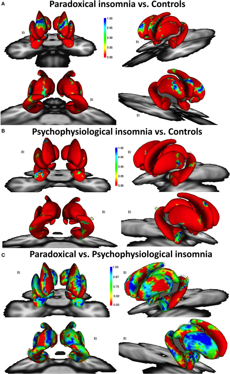
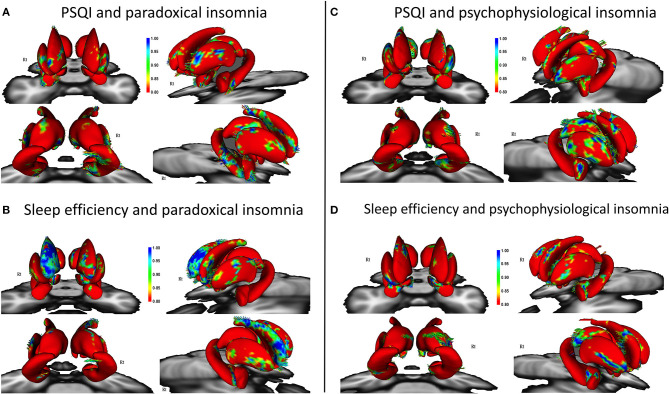
Insomnia disorder (ID) is a common illness associated with mood and cognitive impairments. Subtyping ID is an ongoing debate in sleep medicine, but the underlying mechanisms of each subtype is poorly understood. Growing evidence suggests that subcortical brain structures play the key roles in pathophysiology of ID and its subtypes. Here, we aimed to investigate structural alteration of subcortical regions in patients with two common ID subtypes i.e., paradoxical and psychophysiological insomnia. Fifty-five patients and 49 healthy controls were recruited for this study and T1-weighted images and subjective and objective sleep parameters (i.e., Pittsburgh Sleep Quality Index and polysomnography) were collected from participants. Subcortical structures including the hippocampus, amygdala, caudate, putamen, globus pallidus, nucleus accumbens, and thalamus were automatically segmented in FSL. Volume and shape (using surface vertices) of each structure were compared between the groups, controlled for covariates, and corrected for multiple comparisons. In addition, correlations of sleep parameters and surface vertices or volumes were calculated. The caudate's volume was smaller in patients than controls. Compared with controls, we found regional shrinkage in the caudate, nucleus accumbens, posterior putamen, hippocampus, thalamus, and amygdala in paradoxical insomnia and shrinkage in the amygdala, caudate, hippocampus, and putamen in psychophysiological insomnia. Interestingly, comparing two patients groups, shape alteration in the caudate, putamen, and nucleus accumbens in paradoxical insomnia and shrinkage in the thalamus, amygdala, and hippocampus in psychophysiological insomnia were observed. Both subjective and objective sleep parameters were associated with these regional shape alterations in patients. Our results support the differential role of subcortical brain structures in pathophysiology of paradoxical and psychophysiological insomnia.
失眠症(ID)是一种与情绪和认知障碍相关的常见疾病。对失眠症进行亚型分类是睡眠医学中一个持续争论的话题,但对每种亚型的潜在机制了解甚少。越来越多的证据表明,皮层下脑结构在失眠症及其亚型的病理生理学中起关键作用。在此,我们旨在研究两种常见失眠症亚型即矛盾性失眠和心理生理性失眠患者皮层下区域的结构改变。本研究招募了55名患者和49名健康对照者,并从参与者处收集了T1加权图像以及主观和客观睡眠参数(即匹兹堡睡眠质量指数和多导睡眠图)。在FSL中自动分割包括海马体、杏仁核、尾状核、壳核、苍白球、伏隔核和丘脑在内的皮层下结构。比较两组之间每种结构的体积和形状(使用表面顶点),对协变量进行控制,并对多重比较进行校正。此外,计算睡眠参数与表面顶点或体积之间的相关性。患者的尾状核体积小于对照组。与对照组相比,我们发现矛盾性失眠患者的尾状核、伏隔核、壳核后部、海马体、丘脑和杏仁核区域萎缩,心理生理性失眠患者的杏仁核、尾状核、海马体和壳核萎缩。有趣的是,比较两组患者,观察到矛盾性失眠患者的尾状核、壳核和伏隔核形状改变,心理生理性失眠患者的丘脑、杏仁核和海马体萎缩。主观和客观睡眠参数均与患者这些区域形状改变相关。我们的结果支持皮层下脑结构在矛盾性失眠和心理生理性失眠病理生理学中的不同作用。