• 文献检索
  • 文档翻译
  • 深度研究
  • 学术资讯
  • Suppr Zotero 插件Zotero 插件
  • 邀请有礼
  • 套餐&价格
  • 历史记录
应用&插件
Suppr Zotero 插件Zotero 插件浏览器插件Mac 客户端Windows 客户端微信小程序
定价
高级版会员购买积分包购买API积分包
服务
文献检索文档翻译深度研究API 文档MCP 服务
关于我们
关于 Suppr公司介绍联系我们用户协议隐私条款
关注我们

Suppr 超能文献

核心技术专利:CN118964589B侵权必究
粤ICP备2023148730 号-1Suppr @ 2026

文献检索

告别复杂PubMed语法,用中文像聊天一样搜索,搜遍4000万医学文献。AI智能推荐,让科研检索更轻松。

立即免费搜索

文件翻译

保留排版,准确专业,支持PDF/Word/PPT等文件格式,支持 12+语言互译。

免费翻译文档

深度研究

AI帮你快速写综述,25分钟生成高质量综述,智能提取关键信息,辅助科研写作。

立即免费体验

基于光学相干断层扫描血管造影的抗血管内皮生长因子治疗期间活动性近视性脉络膜新生血管形态学变化的定量评估

Optical Coherence Tomography Angiography-Based Quantitative Assessment of Morphologic Changes in Active Myopic Choroidal Neovascularization During Anti-vascular Endothelial Growth Factor Therapy.

作者信息

Wang Yao, Hu Zhongli, Zhu Tiepei, Su Zhitao, Fang Xiaoyun, Lin Jijian, Chen Zhiqing, Su Zhaoan, Ye Panpan, Ma Jian, Zhang Li, Li Jinyu, Feng Lei, Sun Chuan-Bin, Zhang Zhiyong, Shentu Xingchao

机构信息

Eye Center of the Second Affiliated Hospital, School of Medicine, Zhejiang University, Hangzhou, China.

Department of Ophthalmology, Zhuji People's Hospital of Zhejiang Province, Zhuji, China.

出版信息

Front Med (Lausanne). 2021 May 7;8:657772. doi: 10.3389/fmed.2021.657772. eCollection 2021.

DOI:10.3389/fmed.2021.657772
PMID:34026789
原文链接:https://pmc.ncbi.nlm.nih.gov/articles/PMC8137977/
Abstract

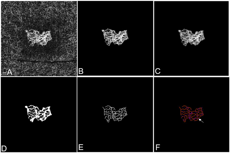
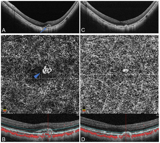
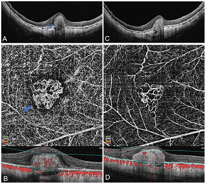
To establish quantitative profile of the morphologic changes among patients with active myopic choroidal neovascularization (mCNV) before and after anti-vascular endothelial growth factor (VEGF) therapy using optical coherence tomography angiography (OCTA) to assess the therapeutic response. Patients with active mCNV who received anti-VEGF injections between February 2017 to October 2020 and fit the study criteria were retrospectively reviewed. Quantitative analysis of their OCTA images were carried out to evaluate the morphologic features and vascular changes of mCNV lesions in response to anti-VEGF therapy. For further quantitative profiling, mCNV area, fractal dimension, vessel area, vessel density, vessel diameter, vessel length, vessel junction, junction density, and vessel tortuosity were obtained by means of advanced skeletonization postprocessing analyses. Thirty-one eyes of 29 consecutive patients with OCTA-positive mCNV lesions (mean spherical equivalent: -12.55 ± 3.24 diopters) were included. The 31 cases were divided into two phenotypes at baseline: organized interlacing pattern (83.87%) and disorganized vascular loops pattern (16.13%). The values of mCNV area, fractal dimension, vessel area, vessel length, vessel junction, and junction density decreased remarkably 1 month after the initial anti-VEGF injection ( < 0.001). Although, vessel density, vessel diameter, and vessel tortuosity increased meanwhile, only vessel diameter displayed statistical significance ( = 0.027). Of note, relative ratio analysis showed that vessel junction was the most sensitive biomarker in response to anti-VEGF therapy, reflecting a mean decrease of 50.36%. Sensitivity lowered successively in biomarkers of vessel length, vessel area, junction density, mCNV area, and fractal dimension. In addition, percent change of mCNV area ( = 0.552, = 0.002), fractal dimension ( = 0.446, = 0.017), vessel area ( = 0.518, = 0.005), and vessel length ( = 0.440, = 0.019) were moderately associated with that of central retinal thickness. The study showed morphological as well as quantitative changes on OCTA responding to anti-VEGF treatment in mCNV patients, among which vessel junctions might be the most predictive biomarker. OCTA-based analysis, providing intuitive images and a large spectrum of quantitative data at the same time, could promote new insights into the therapeutic response assessment in mCNV patients.

摘要

利用光学相干断层扫描血管造影(OCTA)建立活动性近视性脉络膜新生血管(mCNV)患者在抗血管内皮生长因子(VEGF)治疗前后形态学变化的定量特征,以评估治疗反应。对2017年2月至2020年10月期间接受抗VEGF注射且符合研究标准的活动性mCNV患者进行回顾性分析。对其OCTA图像进行定量分析,以评估抗VEGF治疗后mCNV病变的形态学特征和血管变化。为进行进一步的定量分析,通过先进的骨架化后处理分析获得mCNV面积、分形维数、血管面积、血管密度、血管直径、血管长度、血管连接、连接密度和血管迂曲度。纳入29例连续患者的31只OCTA阳性mCNV病变眼(平均球镜等效度:-12.55±3.24屈光度)。31例患者在基线时分为两种表型:有组织的交织模式(83.87%)和无组织的血管环模式(16.13%)。首次抗VEGF注射后1个月,mCNV面积、分形维数、血管面积、血管长度、血管连接和连接密度值显著下降(<0.001)。虽然同时血管密度、血管直径和血管迂曲度增加,但只有血管直径具有统计学意义(=0.027)。值得注意的是,相对比率分析显示,血管连接是对抗VEGF治疗反应最敏感的生物标志物,平均下降50.36%。血管长度、血管面积、连接密度、mCNV面积和分形维数的生物标志物敏感性依次降低。此外,mCNV面积(=0.552,=0.002)、分形维数(=0.446,=0.017)、血管面积(=0.518,=0.005)和血管长度(=0.440,=0.019)的百分比变化与中心视网膜厚度的百分比变化中度相关。该研究显示了mCNV患者抗VEGF治疗后OCTA上的形态学和定量变化,其中血管连接可能是最具预测性的生物标志物。基于OCTA的分析同时提供直观图像和大量定量数据,可促进对mCNV患者治疗反应评估的新认识。

https://cdn.ncbi.nlm.nih.gov/pmc/blobs/4c9d/8137977/160205bc3cf7/fmed-08-657772-g0003.jpg
https://cdn.ncbi.nlm.nih.gov/pmc/blobs/4c9d/8137977/56720949de8b/fmed-08-657772-g0001.jpg
https://cdn.ncbi.nlm.nih.gov/pmc/blobs/4c9d/8137977/2022abab1096/fmed-08-657772-g0002.jpg
https://cdn.ncbi.nlm.nih.gov/pmc/blobs/4c9d/8137977/160205bc3cf7/fmed-08-657772-g0003.jpg
https://cdn.ncbi.nlm.nih.gov/pmc/blobs/4c9d/8137977/56720949de8b/fmed-08-657772-g0001.jpg
https://cdn.ncbi.nlm.nih.gov/pmc/blobs/4c9d/8137977/2022abab1096/fmed-08-657772-g0002.jpg
https://cdn.ncbi.nlm.nih.gov/pmc/blobs/4c9d/8137977/160205bc3cf7/fmed-08-657772-g0003.jpg

相似文献

1
Optical Coherence Tomography Angiography-Based Quantitative Assessment of Morphologic Changes in Active Myopic Choroidal Neovascularization During Anti-vascular Endothelial Growth Factor Therapy.基于光学相干断层扫描血管造影的抗血管内皮生长因子治疗期间活动性近视性脉络膜新生血管形态学变化的定量评估
Front Med (Lausanne). 2021 May 7;8:657772. doi: 10.3389/fmed.2021.657772. eCollection 2021.
2
Macular density alterations in myopic choroidal neovascularization and the effect of anti-VEGF on it.近视性脉络膜新生血管中的黄斑密度改变及其抗VEGF治疗效果
Int J Ophthalmol. 2021 Aug 18;14(8):1205-1212. doi: 10.18240/ijo.2021.08.11. eCollection 2021.
3
PREDICTING LESION SHRINKAGE IN EYES WITH MYOPIC CHOROIDAL NEOVASCULARIZATION FROM FEATURES ON OPTICAL COHERENCE TOMOGRAPHY ANGIOGRAPHY.基于光学相干断层扫描血管造影特征预测近视性脉络膜新生血管眼中病变的缩小情况
Retina. 2022 Sep 1;42(9):1665-1672. doi: 10.1097/IAE.0000000000003526.
4
Novel Predictors of Visual Outcome in Anti-VEGF Therapy for Myopic Choroidal Neovascularization Derived Using OCT Angiography.使用光学相干断层扫描血管造影术得出的近视性脉络膜新生血管抗VEGF治疗视觉结果的新型预测指标
Ophthalmol Retina. 2018 Nov;2(11):1118-1124. doi: 10.1016/j.oret.2018.04.011. Epub 2018 May 31.
5
Perforating scleral vessels adjacent to myopic choroidal neovascularization achieved a poor outcome after intravitreal anti-VEGF therapy.在玻璃体内抗血管内皮生长因子(VEGF)治疗后,近视性脉络膜新生血管附近的巩膜穿孔血管预后较差。
Front Med (Lausanne). 2022 Dec 13;9:1065397. doi: 10.3389/fmed.2022.1065397. eCollection 2022.
6
An ImageJ macro tool for OCTA-based quantitative analysis of Myopic Choroidal neovascularization.基于 OCTA 的近视性脉络膜新生血管定量分析的 ImageJ 宏工具。
PLoS One. 2023 Apr 21;18(4):e0283929. doi: 10.1371/journal.pone.0283929. eCollection 2023.
7
Using optical coherence tomography angiography to guide myopic choroidal neovascularization treatment: a 3-year follow-up study.应用光相干断层扫描血管造影指导近视脉络膜新生血管的治疗:一项 3 年随访研究。
Graefes Arch Clin Exp Ophthalmol. 2021 Nov;259(11):3295-3303. doi: 10.1007/s00417-021-05270-5. Epub 2021 Jun 18.
8
ASSESSING THE ACTIVITY OF MYOPIC CHOROIDAL NEOVASCULARIZATION: Comparison Between Optical Coherence Tomography Angiography and Dye Angiography.评估近视性脉络膜新生血管的活性:光学相干断层扫描血管造影与染料血管造影的比较
Retina. 2020 Sep;40(9):1757-1764. doi: 10.1097/IAE.0000000000002650.
9
Retinal vascular changes and aqueous humor cytokines changes after aflibercept intravitreal injection in treatment-naïve myopic choroidal neovascularization.治疗初发近视性脉络膜新生血管时阿柏西普玻璃体内注射后的视网膜血管变化和房水细胞因子变化。
Sci Rep. 2018 Oct 23;8(1):15631. doi: 10.1038/s41598-018-33926-6.
10
Choroidal Structural Changes in Myopic Choroidal Neovascularization After Treatment With Antivascular Endothelial Growth Factor Over 1 Year.抗血管内皮生长因子治疗1年以上后近视性脉络膜新生血管的脉络膜结构变化
Invest Ophthalmol Vis Sci. 2016 Sep 1;57(11):4933-4939. doi: 10.1167/iovs.16-20191.

引用本文的文献

1
Short-Term Morphological and Quantitative Changes in Non-Exudative Macular Neovascularization Using Spectral-Domain OCT and OCT Angiography: A Pilot Study.使用光谱域光学相干断层扫描(SD-OCT)和光学相干断层扫描血管造影(OCTA)对非渗出性黄斑新生血管进行的短期形态学和定量变化:一项初步研究
J Clin Med. 2025 May 22;14(11):3622. doi: 10.3390/jcm14113622.
2
OCT Angiography of Type 1 Macular Neovascularization in AMD: A Morphometric Evaluation.年龄相关性黄斑变性中1型黄斑新生血管的光学相干断层扫描血管造影:形态学评估
Transl Vis Sci Technol. 2025 Feb 3;14(2):23. doi: 10.1167/tvst.14.2.23.
3
An ImageJ macro tool for OCTA-based quantitative analysis of Myopic Choroidal neovascularization.

本文引用的文献

1
Assessment of conbercept therapy for high myopia macular neovascularization by optical coherence tomography angiography.光学相干断层扫描血管造影评估康柏西普治疗高度近视性黄斑新生血管。
Sci Rep. 2020 Oct 12;10(1):16959. doi: 10.1038/s41598-020-74073-1.
2
Profiling neovascular age-related macular degeneration choroidal neovascularization lesion response to anti-vascular endothelial growth factor therapy using SSOCTA.应用 SSOCTA 对新生血管性年龄相关性黄斑变性脉络膜新生血管病变对抗血管内皮生长因子治疗的反应进行分析。
Acta Ophthalmol. 2021 Mar;99(2):e240-e246. doi: 10.1111/aos.14554. Epub 2020 Jul 24.
3
Prevalence and associations of myopia in Hong Kong primary school students.
基于 OCTA 的近视性脉络膜新生血管定量分析的 ImageJ 宏工具。
PLoS One. 2023 Apr 21;18(4):e0283929. doi: 10.1371/journal.pone.0283929. eCollection 2023.
4
Vascular Aging and Damage in Patients with Iron Metabolism Disorders.铁代谢紊乱患者的血管衰老与损伤
Diagnostics (Basel). 2022 Nov 16;12(11):2817. doi: 10.3390/diagnostics12112817.
5
Changes on optical coherence tomography angiography and fluorescein angiography in eyes with neovascular age-related macular degeneration.新生血管性年龄相关性黄斑变性患者眼部光学相干断层扫描血管造影和荧光素血管造影的变化
Int J Ophthalmol. 2022 Nov 18;15(11):1837-1844. doi: 10.18240/ijo.2022.11.15. eCollection 2022.
6
Advances in OCT Imaging in Myopia and Pathologic Myopia.近视和病理性近视的光学相干断层扫描成像进展
Diagnostics (Basel). 2022 Jun 8;12(6):1418. doi: 10.3390/diagnostics12061418.
7
Performance Assessment of Two Different Approaches of Measuring Skeletonized Radial Peripapillary Capillary Vessel Density in Glaucoma Patients.青光眼患者中两种测量视网膜乳头周围骨小梁化的桡侧毛细血管密度不同方法的性能评估
Front Med (Lausanne). 2022 Mar 16;8:814306. doi: 10.3389/fmed.2021.814306. eCollection 2021.
8
Novel Findings of Retinal and Choroidal Features Utilizing Optical Coherence Tomography Angiography Analysis in Patients With Autoimmune Posterior Uveitis.利用光学相干断层扫描血管造影分析自身免疫性后葡萄膜炎患者视网膜和脉络膜特征的新发现。
Front Med (Lausanne). 2022 Jan 11;8:801036. doi: 10.3389/fmed.2021.801036. eCollection 2021.
香港小学生近视的患病率及其关联因素
Jpn J Ophthalmol. 2020 Jul;64(4):437-449. doi: 10.1007/s10384-020-00733-4. Epub 2020 Mar 16.
4
OCT Angiography Features of Neovascularization as Predictive Factors for Frequent Recurrence in Age-Related Macular Degeneration.OCT 血管造影对年龄相关性黄斑变性新生血管特征的预测作用及其与频繁复发的相关性。
Am J Ophthalmol. 2020 May;213:109-119. doi: 10.1016/j.ajo.2020.01.012. Epub 2020 Jan 16.
5
Microvasculature Changes of Myopic Choroidal Neovascularization and the Predictive Value of Feeder Vessel Disappearance after Ranibizumab Treatment Revealed Using Optical Coherence Tomography Angiography.利用光学相干断层扫描血管造影术观察近视脉络膜新生血管的微血管变化及雷珠单抗治疗后供养血管消失的预测价值。
Ophthalmologica. 2020;243(4):263-270. doi: 10.1159/000504755. Epub 2019 Dec 13.
6
COMPARISON OF PROJECTION-RESOLVED OPTICAL COHERENCE TOMOGRAPHY ANGIOGRAPHY-BASED METRICS FOR THE EARLY DETECTION OF RETINAL MICROVASCULAR IMPAIRMENTS IN DIABETES MELLITUS.基于光相干断层扫描血管造影的投影分辨测量在糖尿病视网膜微血管损伤早期检测中的比较。
Retina. 2020 Sep;40(9):1783-1792. doi: 10.1097/IAE.0000000000002655.
7
IMPACT OF OPTICAL COHERENCE TOMOGRAPHY ANGIOGRAPHY REVIEW STRATEGY ON DETECTION OF CHOROIDAL NEOVASCULARIZATION.光学相干断层扫描血管造影复查策略对脉络膜新生血管检测的影响
Retina. 2020 Apr;40(4):672-678. doi: 10.1097/IAE.0000000000002443.
8
Influence of myopic macular degeneration severity on treatment outcomes with intravitreal aflibercept in the MYRROR study.在 MYRROR 研究中,近视性黄斑变性严重程度对玻璃体内注射阿柏西普治疗效果的影响。
Acta Ophthalmol. 2019 Aug;97(5):e729-e735. doi: 10.1111/aos.14035. Epub 2019 Jan 28.
9
Repeatability and reliability of quantitative fundus autofluorescence imaging in patients with early and intermediate age-related macular degeneration.定量眼底自发荧光成像在早期和中期年龄相关性黄斑变性患者中的重复性和可靠性。
Acta Ophthalmol. 2019 Jun;97(4):e526-e532. doi: 10.1111/aos.13987. Epub 2018 Dec 13.
10
Vascular remodeling of choroidal neovascularization in older myopic patients treated with ranibizumab.雷珠单抗治疗老年近视患者脉络膜新生血管的血管重塑
Graefes Arch Clin Exp Ophthalmol. 2019 Mar;257(3):485-493. doi: 10.1007/s00417-018-04205-x. Epub 2018 Dec 7.