Dong Li, Yang Qiong, Zhang Rui Heng, Wei Wen Bin
Beijing Tongren Eye Center, Beijing Key Laboratory of Intraocular Tumor Diagnosis and Treatment, Beijing Ophthalmology & Visual Sciences Key Lab, Medical Artificial Intelligence Research and Verification Key Laboratory of the Ministry of Industry and Information Technology, Beijing Tongren Hospital, Capital Medical University, 1 Dong Jiao Min Lane, Beijing 100730, China.
EClinicalMedicine. 2021 May 8;35:100875. doi: 10.1016/j.eclinm.2021.100875. eCollection 2021 May.
Age-related macular degeneration (AMD) is one of the leading causes of vision loss in the elderly population. The application of artificial intelligence (AI) provides convenience for the diagnosis of AMD. This systematic review and meta-analysis aimed to quantify the performance of AI in detecting AMD in fundus photographs.
We searched PubMed, Embase, Web of Science and the Cochrane Library before December 31st, 2020 for studies reporting the application of AI in detecting AMD in color fundus photographs. Then, we pooled the data for analysis. PROSPERO registration number: CRD42020197532.
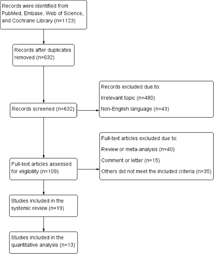
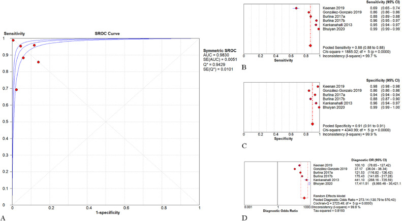
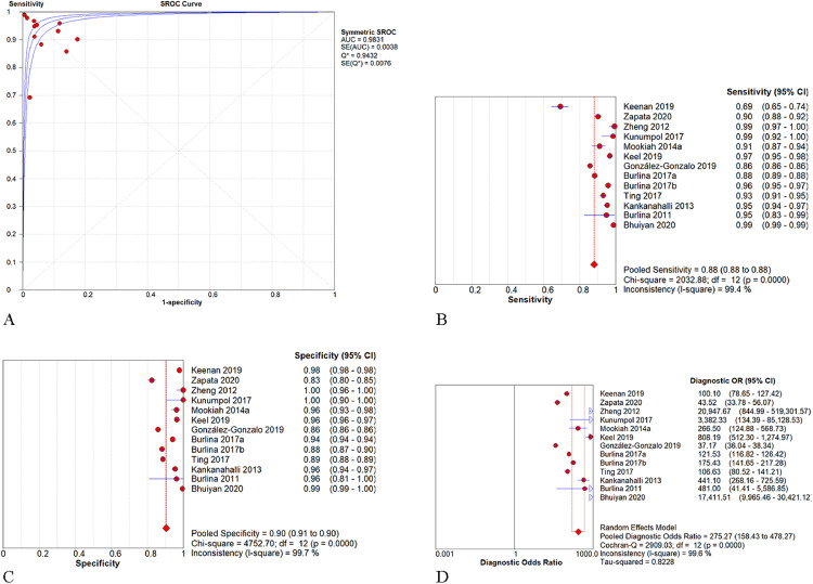
19 studies were finally selected for systematic review and 13 of them were included in the quantitative synthesis. All studies adopted human graders as reference standard. The pooled area under the receiver operating characteristic curve (AUROC) was 0.983 (95% confidence interval (CI):0.979-0.987). The pooled sensitivity, specificity, and diagnostic odds ratio (DOR) were 0.88 (95% CI:0.88-0.88), 0.90 (95% CI:0.90-0.91), and 275.27 (95% CI:158.43-478.27), respectively. Threshold analysis was performed and a potential threshold effect was detected among the studies (Spearman correlation coefficient: -0.600, = 0.030), which was the main cause for the heterogeneity. For studies applying convolutional neural networks in the Age-Related Eye Disease Study database, the pooled AUROC, sensitivity, specificity, and DOR were 0.983 (95% CI:0.978-0.988), 0.88 (95% CI:0.88-0.88), 0.91 (95% CI:0.91-0.91), and 273.14 (95% CI:130.79-570.43), respectively.
Our data indicated that AI was able to detect AMD in color fundus photographs. The application of AI-based automatic tools is beneficial for the diagnosis of AMD.
Capital Health Research and Development of Special (2020-1-2052).
年龄相关性黄斑变性(AMD)是老年人群视力丧失的主要原因之一。人工智能(AI)的应用为AMD的诊断提供了便利。本系统评价和荟萃分析旨在量化AI在眼底照片中检测AMD的性能。
我们在2020年12月31日前检索了PubMed、Embase、Web of Science和Cochrane图书馆,以查找报告AI在彩色眼底照片中检测AMD应用的研究。然后,我们汇总数据进行分析。PROSPERO注册号:CRD42020197532。
最终选择19项研究进行系统评价,其中13项纳入定量合成。所有研究均采用人工分级作为参考标准。汇总的受试者工作特征曲线下面积(AUROC)为0.983(95%置信区间(CI):0.979-0.987)。汇总的敏感性、特异性和诊断比值比(DOR)分别为0.88(95%CI:0.88-0.88)、0.90(95%CI:0.90-0.91)和275.27(95%CI:158.43-478.27)。进行了阈值分析,在研究中检测到潜在的阈值效应(Spearman相关系数:-0.600,P = 0.030),这是异质性的主要原因。对于在年龄相关性眼病研究数据库中应用卷积神经网络的研究,汇总的AUROC、敏感性、特异性和DOR分别为0.983(95%CI:0.978-0.988)、0.88(95%CI:0.88-0.88)、0.91(95%CI:0.91-0.91)和273.14(95%CI:130.79-570.43)。
我们的数据表明,AI能够在彩色眼底照片中检测AMD。基于AI的自动工具的应用有利于AMD的诊断。
首都卫生发展科研专项(2020-1-2052)。