Liu Xin-Yuan, Song Wen, Mao Tao, Zhang Qi, Zhang Cuiping, Li Xiao-Yu
Department of Gastroenterology, The Affiliated Hospital of Qingdao University, Qingdao, China.
Front Oncol. 2022 Aug 15;12:915481. doi: 10.3389/fonc.2022.915481. eCollection 2022.
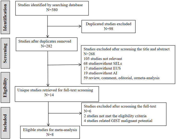
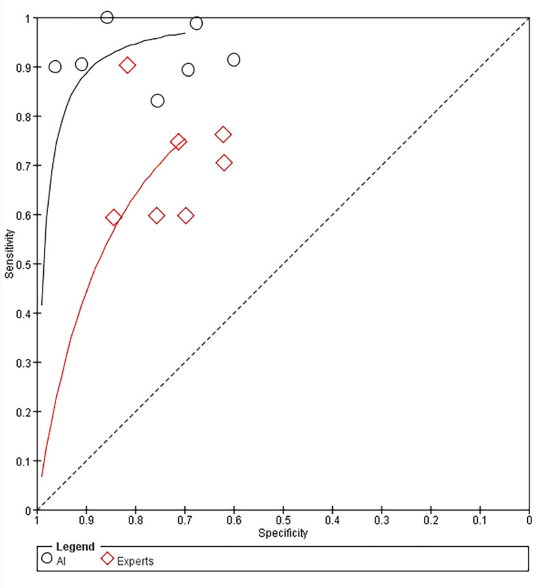
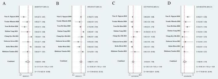
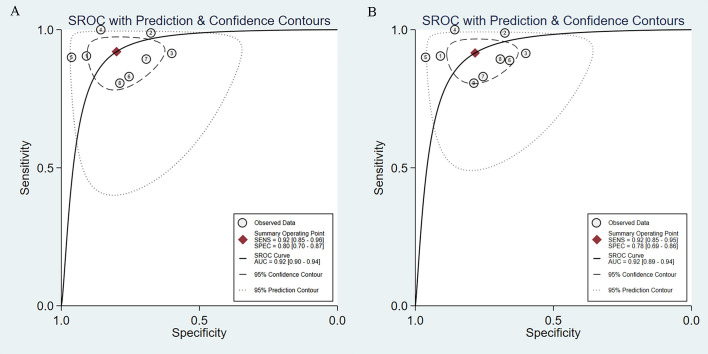
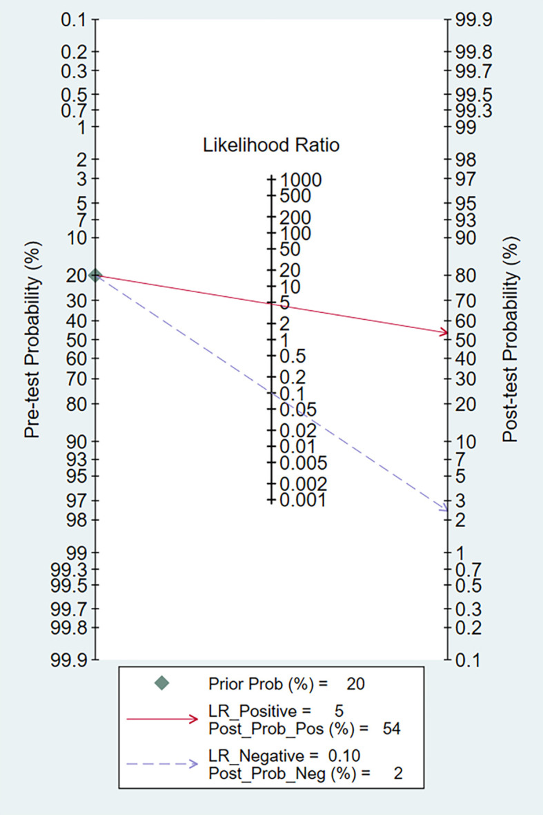
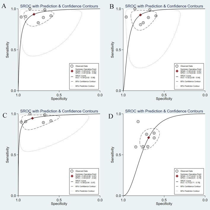
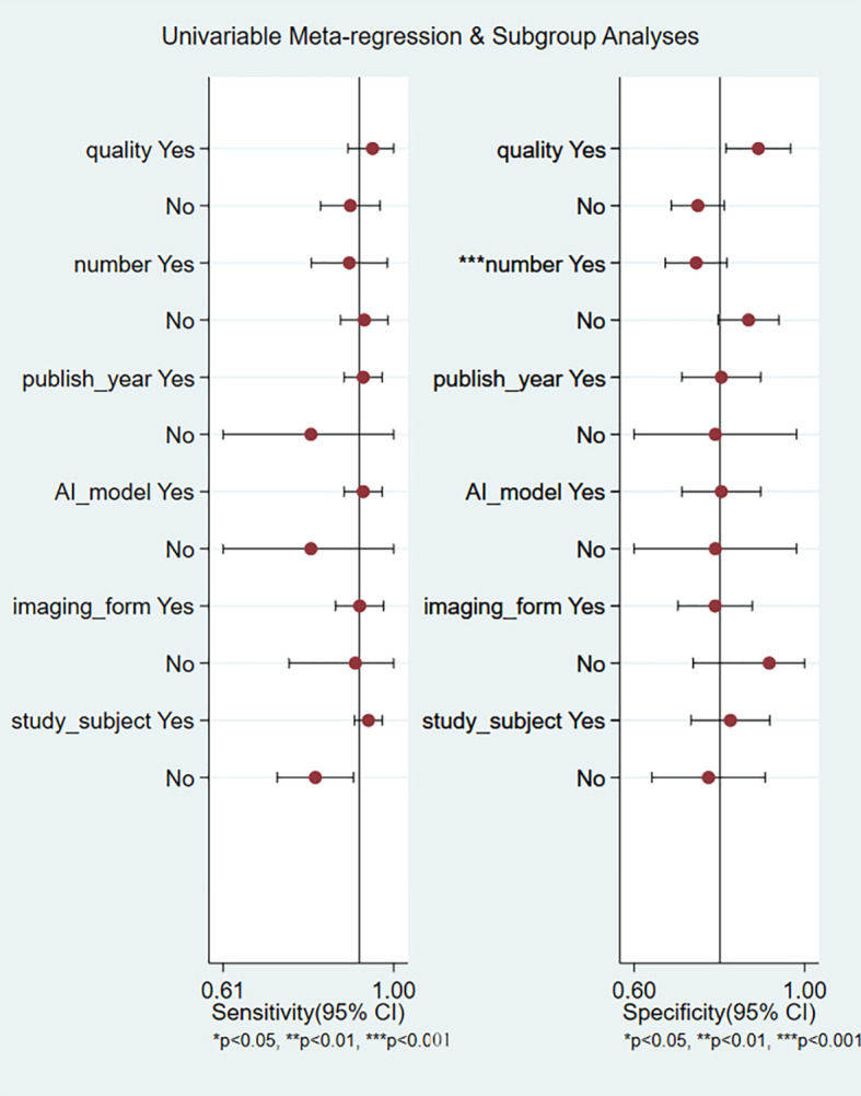
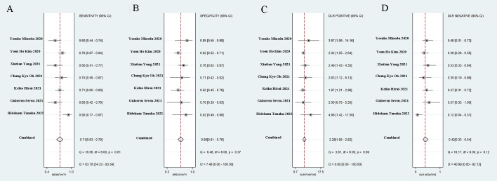
Endoscopic ultrasonography (EUS) is the most common method for diagnosing gastrointestinal subepithelial lesions (SELs); however, it usually requires histopathological confirmation using invasive methods. Artificial intelligence (AI) algorithms have made significant progress in medical imaging diagnosis. The purpose of our research was to explore the application of AI in the diagnosis of SELs using EUS and to evaluate the diagnostic performance of AI-assisted EUS. Three databases, PubMed, EMBASE, and the Cochrane Library, were comprehensively searched for relevant literature. RevMan 5.4.1 and Stata 17.0, were used to calculate and analyze the combined sensitivity, specificity, positive likelihood ratio (PLR), negative likelihood ratio (NLR), diagnostic odds ratio (DOR), and summary receiver-operating characteristic curve (SROC). Eight studies were selected from 380 potentially relevant studies for the meta-analysis of AI-aided EUS diagnosis of SELs. The combined sensitivity, specificity, and DOR of AI-aided EUS were 0.92 (95% CI, 0.85-0.96), 0.80 (95% CI, 0.70-0.87), and 46.27 (95% CI, 19.36-110.59), respectively). The area under the curve (AUC) was 0.92 (95% CI, 0.90-0.94). The AI model in differentiating GIST from leiomyoma had a pooled AUC of 0.95, sensitivity of 0.93, specificity of 0.88, PLR of 8.04, and NLR of 0.08. The combined sensitivity, specificity, and AUC of the AI-aided EUS diagnosis in the convolutional neural network (CNN) model were 0.93, 0.81, and 0.94, respectively. AI-aided EUS diagnosis using conventional brightness mode (B-mode) EUS images had a combined sensitivity of 0.92, specificity of 0.79, and AUC of 0.92. AI-aided EUS diagnosis based on patients had a combined sensitivity, specificity, and AUC of 0.95, 0.83, and 0.96, respectively. Additionally, AI-aided EUS was superior to EUS by experts in terms of sensitivity (0.93 vs. 0.71), specificity (0.81 vs. 0.69), and AUC (0.94 vs. 0.75). In conclusion, AI-assisted EUS is a promising and reliable method for distinguishing SELs, with excellent diagnostic performance. More multicenter cohort and prospective studies are expected to be conducted to further develop AI-assisted real-time diagnostic systems and validate the superiority of AI systems. PROSPERO (https://www.crd.york.ac.uk/PROSPERO/), identifier CRD42022303990.
内镜超声检查(EUS)是诊断胃肠道上皮下病变(SELs)最常用的方法;然而,它通常需要使用侵入性方法进行组织病理学确认。人工智能(AI)算法在医学影像诊断方面取得了重大进展。我们研究的目的是探索AI在使用EUS诊断SELs中的应用,并评估AI辅助EUS的诊断性能。全面检索了三个数据库,即PubMed、EMBASE和Cochrane图书馆,以获取相关文献。使用RevMan 5.4.1和Stata 17.0计算并分析合并敏感度、特异度、阳性似然比(PLR)、阴性似然比(NLR)、诊断比值比(DOR)和汇总接受者操作特征曲线(SROC)。从380项潜在相关研究中筛选出8项研究,用于AI辅助EUS诊断SELs的荟萃分析。AI辅助EUS的合并敏感度、特异度和DOR分别为0.92(95%CI,0.85-0.96)、0.80(95%CI,0.70-0.87)和46.27(95%CI,19.36-110.59)。曲线下面积(AUC)为0.92(95%CI,0.90-0.94)。AI模型在区分胃肠道间质瘤(GIST)和平滑肌瘤方面的合并AUC为0.95,敏感度为0.93,特异度为0.88,PLR为8.04,NLR为0.08。卷积神经网络(CNN)模型中AI辅助EUS诊断的合并敏感度、特异度和AUC分别为0.93、0.81和0.94。使用传统亮度模式(B模式)EUS图像进行AI辅助EUS诊断的合并敏感度为0.92,特异度为0.79,AUC为0.92。基于患者的AI辅助EUS诊断的合并敏感度、特异度和AUC分别为0.95、0.83和0.96。此外,在敏感度(0.93对0.71)、特异度(0.81对0.69)和AUC(0.94对0.75)方面,AI辅助EUS优于专家进行的EUS。总之,AI辅助EUS是一种有前景且可靠的区分SELs的方法,具有出色的诊断性能。预计将开展更多的多中心队列研究和前瞻性研究,以进一步开发AI辅助实时诊断系统并验证AI系统的优越性。PROSPERO(https://www.crd.york.ac.uk/PROSPERO/),标识符CRD42022303990。