• 文献检索
  • 文档翻译
  • 深度研究
  • 学术资讯
  • Suppr Zotero 插件Zotero 插件
  • 邀请有礼
  • 套餐&价格
  • 历史记录
应用&插件
Suppr Zotero 插件Zotero 插件浏览器插件Mac 客户端Windows 客户端微信小程序
定价
高级版会员购买积分包购买API积分包
服务
文献检索文档翻译深度研究API 文档MCP 服务
关于我们
关于 Suppr公司介绍联系我们用户协议隐私条款
关注我们

Suppr 超能文献

核心技术专利:CN118964589B侵权必究
粤ICP备2023148730 号-1Suppr @ 2026

文献检索

告别复杂PubMed语法,用中文像聊天一样搜索,搜遍4000万医学文献。AI智能推荐,让科研检索更轻松。

立即免费搜索

文件翻译

保留排版,准确专业,支持PDF/Word/PPT等文件格式,支持 12+语言互译。

免费翻译文档

深度研究

AI帮你快速写综述,25分钟生成高质量综述,智能提取关键信息,辅助科研写作。

立即免费体验

深度学习方法自动检测彩色眼底照片中的地理萎缩。

A Deep Learning Approach for Automated Detection of Geographic Atrophy from Color Fundus Photographs.

机构信息

Division of Epidemiology and Clinical Applications, National Eye Institute, National Institutes of Health, Bethesda, Maryland.

Division of Epidemiology and Clinical Applications, National Eye Institute, National Institutes of Health, Bethesda, Maryland; National Center for Biotechnology Information, National Library of Medicine, National Institutes of Health, Bethesda, Maryland.

出版信息

Ophthalmology. 2019 Nov;126(11):1533-1540. doi: 10.1016/j.ophtha.2019.06.005. Epub 2019 Jun 11.

DOI:10.1016/j.ophtha.2019.06.005
PMID:31358385
原文链接:https://pmc.ncbi.nlm.nih.gov/articles/PMC6810830/
Abstract

PURPOSE

To assess the utility of deep learning in the detection of geographic atrophy (GA) from color fundus photographs and to explore potential utility in detecting central GA (CGA).

DESIGN

A deep learning model was developed to detect the presence of GA in color fundus photographs, and 2 additional models were developed to detect CGA in different scenarios.

PARTICIPANTS

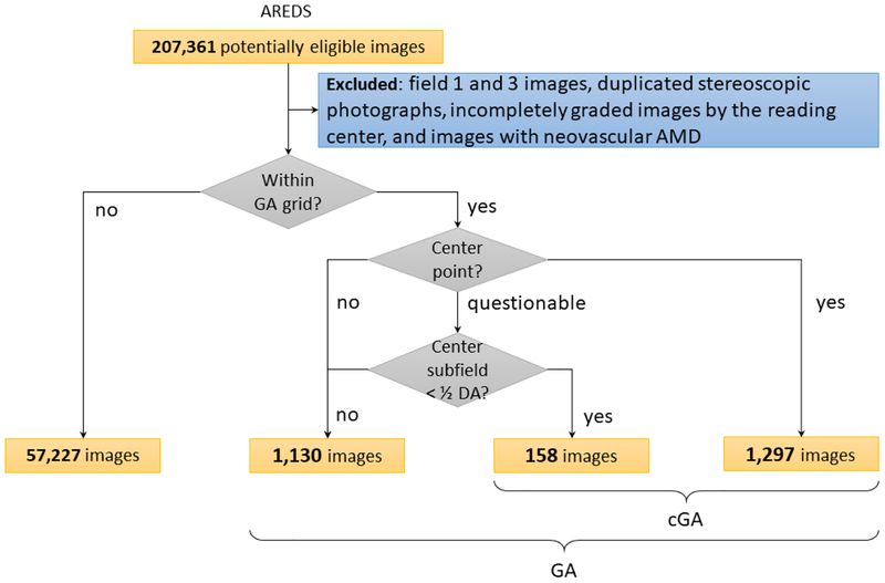
A total of 59 812 color fundus photographs from longitudinal follow-up of 4582 participants in the Age-Related Eye Disease Study (AREDS) dataset. Gold standard labels were from human expert reading center graders using a standardized protocol.

METHODS

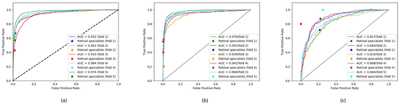
A deep learning model was trained to use color fundus photographs to predict GA presence from a population of eyes with no AMD to advanced AMD. A second model was trained to predict CGA presence from the same population. A third model was trained to predict CGA presence from the subset of eyes with GA. For training and testing, 5-fold cross-validation was used. For comparison with human clinician performance, model performance was compared with that of 88 retinal specialists.

MAIN OUTCOME MEASURES

Area under the curve (AUC), accuracy, sensitivity, specificity, and precision.

RESULTS

The deep learning models (GA detection, CGA detection from all eyes, and centrality detection from GA eyes) had AUCs of 0.933-0.976, 0.939-0.976, and 0.827-0.888, respectively. The GA detection model had accuracy, sensitivity, specificity, and precision of 0.965 (95% confidence interval [CI], 0.959-0.971), 0.692 (0.560-0.825), 0.978 (0.970-0.985), and 0.584 (0.491-0.676), respectively, compared with 0.975 (0.971-0.980), 0.588 (0.468-0.707), 0.982 (0.978-0.985), and 0.368 (0.230-0.505) for the retinal specialists. The CGA detection model had values of 0.966 (0.957-0.975), 0.763 (0.641-0.885), 0.971 (0.960-0.982), and 0.394 (0.341-0.448). The centrality detection model had values of 0.762 (0.725-0.799), 0.782 (0.618-0.945), 0.729 (0.543-0.916), and 0.799 (0.710-0.888).

CONCLUSIONS

A deep learning model demonstrated high accuracy for the automated detection of GA. The AUC was noninferior to that of human retinal specialists. Deep learning approaches may also be applied to the identification of CGA. The code and pretrained models are publicly available at https://github.com/ncbi-nlp/DeepSeeNet.

摘要

目的

评估深度学习在检测彩色眼底照片中的地图状萎缩(GA)中的应用,并探索其在检测中心性 GA(CGA)中的潜在应用。

设计

开发了一种深度学习模型来检测彩色眼底照片中 GA 的存在,并开发了另外两种模型来在不同场景中检测 CGA。

参与者

来自年龄相关性眼病研究(AREDS)数据集的 4582 名参与者的纵向随访共 59812 张彩色眼底照片。金标准标签是由人类专家阅读中心分级器使用标准化协议获得的。

方法

训练深度学习模型使用彩色眼底照片从无 AMD 至晚期 AMD 的人群中预测 GA 的存在。第二个模型用于从相同人群中预测 CGA 的存在。第三个模型用于从有 GA 的眼中预测 CGA 的存在。用于训练和测试的是 5 折交叉验证。为了与人类临床医生的表现进行比较,比较了模型性能与 88 名视网膜专家的表现。

主要观察指标

曲线下面积(AUC)、准确性、灵敏度、特异性和精度。

结果

深度学习模型(GA 检测、来自所有眼睛的 CGA 检测和来自 GA 眼睛的中心性检测)的 AUC 分别为 0.933-0.976、0.939-0.976 和 0.827-0.888。GA 检测模型的准确性、灵敏度、特异性和精度分别为 0.965(95%置信区间[CI],0.959-0.971)、0.692(0.560-0.825)、0.978(0.970-0.985)和 0.584(0.491-0.676),而视网膜专家的准确性、灵敏度、特异性和精度分别为 0.975(0.971-0.980)、0.588(0.468-0.707)、0.982(0.978-0.985)和 0.368(0.230-0.505)。CGA 检测模型的数值为 0.966(0.957-0.975)、0.763(0.641-0.885)、0.971(0.960-0.982)和 0.394(0.341-0.448)。中心性检测模型的数值为 0.762(0.725-0.799)、0.782(0.618-0.945)、0.729(0.543-0.916)和 0.799(0.710-0.888)。

结论

深度学习模型在 GA 的自动检测中表现出很高的准确性。AUC 与人类视网膜专家相当。深度学习方法也可用于 CGA 的识别。代码和预训练模型可在 https://github.com/ncbi-nlp/DeepSeeNet 上获得。

https://cdn.ncbi.nlm.nih.gov/pmc/blobs/8973/6810830/bcb00169017d/nihms-1536372-f0002.jpg
https://cdn.ncbi.nlm.nih.gov/pmc/blobs/8973/6810830/ec0e6e56ad2c/nihms-1536372-f0001.jpg
https://cdn.ncbi.nlm.nih.gov/pmc/blobs/8973/6810830/bcb00169017d/nihms-1536372-f0002.jpg
https://cdn.ncbi.nlm.nih.gov/pmc/blobs/8973/6810830/ec0e6e56ad2c/nihms-1536372-f0001.jpg
https://cdn.ncbi.nlm.nih.gov/pmc/blobs/8973/6810830/bcb00169017d/nihms-1536372-f0002.jpg

相似文献

1
A Deep Learning Approach for Automated Detection of Geographic Atrophy from Color Fundus Photographs.深度学习方法自动检测彩色眼底照片中的地理萎缩。
Ophthalmology. 2019 Nov;126(11):1533-1540. doi: 10.1016/j.ophtha.2019.06.005. Epub 2019 Jun 11.
2
DeepSeeNet: A Deep Learning Model for Automated Classification of Patient-based Age-related Macular Degeneration Severity from Color Fundus Photographs.深见网络:一种基于深度学习的用于自动分类基于患者的年龄相关性黄斑变性严重程度的彩色眼底照片的模型。
Ophthalmology. 2019 Apr;126(4):565-575. doi: 10.1016/j.ophtha.2018.11.015. Epub 2018 Nov 22.
3
Deep Learning Automated Detection of Reticular Pseudodrusen from Fundus Autofluorescence Images or Color Fundus Photographs in AREDS2.深度学习自动检测 AREDS2 中的眼底自发荧光图像或彩色眼底照片中的网状假性色素沉着
Ophthalmology. 2020 Dec;127(12):1674-1687. doi: 10.1016/j.ophtha.2020.05.036. Epub 2020 May 21.
4
Evaluation of Geographic Atrophy from Color Photographs and Fundus Autofluorescence Images: Age-Related Eye Disease Study 2 Report Number 11.基于彩色照片和眼底自发荧光图像对地图样萎缩的评估:年龄相关性眼病研究2报告编号11
Ophthalmology. 2016 Nov;123(11):2401-2407. doi: 10.1016/j.ophtha.2016.06.025. Epub 2016 Jul 19.
5
Deep learning-based automated detection of glaucomatous optic neuropathy on color fundus photographs.基于深度学习的彩色眼底照片中青光眼视神经病变的自动检测。
Graefes Arch Clin Exp Ophthalmol. 2020 Apr;258(4):851-867. doi: 10.1007/s00417-020-04609-8. Epub 2020 Jan 27.
6
Deep learning-based detection and classification of geographic atrophy using a deep convolutional neural network classifier.使用深度卷积神经网络分类器基于深度学习的地理性萎缩检测与分类
Graefes Arch Clin Exp Ophthalmol. 2018 Nov;256(11):2053-2060. doi: 10.1007/s00417-018-4098-2. Epub 2018 Aug 8.
7
A Deep Learning Algorithm for Prediction of Age-Related Eye Disease Study Severity Scale for Age-Related Macular Degeneration from Color Fundus Photography.一种基于深度学习的算法,可从眼底彩色照相图预测年龄相关性眼病研究严重程度评分-年龄相关性黄斑变性。
Ophthalmology. 2018 Sep;125(9):1410-1420. doi: 10.1016/j.ophtha.2018.02.037. Epub 2018 Apr 10.
8
Automated image alignment and segmentation to follow progression of geographic atrophy in age-related macular degeneration.自动图像配准与分割以跟踪年龄相关性黄斑变性中地图样萎缩的进展情况。
Retina. 2014 Jul;34(7):1296-307. doi: 10.1097/IAE.0000000000000069.
9
Efficacy of a Deep Learning System for Detecting Glaucomatous Optic Neuropathy Based on Color Fundus Photographs.基于眼底彩色照片的深度学习系统检测青光眼视神经病变的效果。
Ophthalmology. 2018 Aug;125(8):1199-1206. doi: 10.1016/j.ophtha.2018.01.023. Epub 2018 Mar 2.
10
Automated segmentation of geographic atrophy of the retinal epithelium via random forests in AREDS color fundus images.通过随机森林对年龄相关性眼病研究(AREDS)彩色眼底图像中的视网膜色素上皮地图样萎缩进行自动分割
Comput Biol Med. 2015 Oct 1;65:124-36. doi: 10.1016/j.compbiomed.2015.06.018. Epub 2015 Jul 9.

引用本文的文献

1
Artificial intelligence-based apps for screening and diagnosing diabetic retinopathy and common ocular disorders.用于筛查和诊断糖尿病视网膜病变及常见眼部疾病的人工智能应用程序。
World J Methodol. 2025 Dec 20;15(4):107166. doi: 10.5662/wjm.v15.i4.107166.
2
Can artificial intelligence with multimodal imaging outperform traditional methods in predicting age-related macular degeneration progression? A systematic review and exploratory meta-analysis.具有多模态成像的人工智能在预测年龄相关性黄斑变性进展方面能否优于传统方法?一项系统评价和探索性荟萃分析。
BMC Med Inform Decis Mak. 2025 Sep 1;25(1):321. doi: 10.1186/s12911-025-03119-z.
3

本文引用的文献

1
A multi-task deep learning model for the classification of Age-related Macular Degeneration.一种用于年龄相关性黄斑变性分类的多任务深度学习模型。
AMIA Jt Summits Transl Sci Proc. 2019 May 6;2019:505-514. eCollection 2019.
2
Assessment of Deep Generative Models for High-Resolution Synthetic Retinal Image Generation of Age-Related Macular Degeneration.评估深度生成模型在年龄相关性黄斑变性高分辨率合成视网膜图像生成中的应用。
JAMA Ophthalmol. 2019 Mar 1;137(3):258-264. doi: 10.1001/jamaophthalmol.2018.6156.
3
DeepSeeNet: A Deep Learning Model for Automated Classification of Patient-based Age-related Macular Degeneration Severity from Color Fundus Photographs.
Deep Learning for Comprehensive Analysis of Retinal Fundus Images: Detection of Systemic and Ocular Conditions.
用于视网膜眼底图像综合分析的深度学习:全身和眼部疾病的检测
Bioengineering (Basel). 2025 Aug 3;12(8):840. doi: 10.3390/bioengineering12080840.
4
Empowering Portable Age-Related Macular Degeneration Screening: Evaluation of a Deep Learning Algorithm for a Smartphone Fundus Camera.赋能便携式年龄相关性黄斑变性筛查:智能手机眼底相机的深度学习算法评估。
BMJ Open. 2024 Sep 5;14(9):e081398. doi: 10.1136/bmjopen-2023-081398.
5
Artificial intelligence and machine learning a new frontier in the diagnosis of ocular adnexal tumors: A review.人工智能与机器学习:眼附属器肿瘤诊断的新前沿:综述
SAGE Open Med. 2024 Aug 27;12:20503121241274197. doi: 10.1177/20503121241274197. eCollection 2024.
6
Measuring Geographic Atrophy Area Using Column-Based Machine Learning Software on Spectral-Domain Optical Coherence Tomography versus Fundus Auto Fluorescence.在频域光学相干断层扫描与眼底自发荧光图像上,使用基于列的机器学习软件测量地理萎缩面积
Bioengineering (Basel). 2024 Aug 19;11(8):849. doi: 10.3390/bioengineering11080849.
7
[Use of artificial intelligence in geographic atrophy in age-related macular degeneration].[人工智能在年龄相关性黄斑变性地理萎缩中的应用]
Ophthalmologie. 2024 Aug;121(8):616-622. doi: 10.1007/s00347-024-02080-y. Epub 2024 Jul 31.
8
Quantifying Geographic Atrophy in Age-Related Macular Degeneration: A Comparative Analysis Across 12 Deep Learning Models.量化年龄相关性黄斑变性中的地理萎缩:12种深度学习模型的比较分析
Invest Ophthalmol Vis Sci. 2024 Jul 1;65(8):42. doi: 10.1167/iovs.65.8.42.
9
Strong versus Weak Data Labeling for Artificial Intelligence Algorithms in the Measurement of Geographic Atrophy.在地理萎缩测量中人工智能算法的强数据标注与弱数据标注对比
Ophthalmol Sci. 2024 Jan 26;4(5):100477. doi: 10.1016/j.xops.2024.100477. eCollection 2024 Sep-Oct.
10
Deep Learning Approaches for Detecting of Nascent Geographic Atrophy in Age-Related Macular Degeneration.用于检测年龄相关性黄斑变性中新生地图样萎缩的深度学习方法
Ophthalmol Sci. 2023 Nov 17;4(3):100428. doi: 10.1016/j.xops.2023.100428. eCollection 2024 May-Jun.
深见网络:一种基于深度学习的用于自动分类基于患者的年龄相关性黄斑变性严重程度的彩色眼底照片的模型。
Ophthalmology. 2019 Apr;126(4):565-575. doi: 10.1016/j.ophtha.2018.11.015. Epub 2018 Nov 22.
4
Use of Deep Learning for Detailed Severity Characterization and Estimation of 5-Year Risk Among Patients With Age-Related Macular Degeneration.利用深度学习对年龄相关性黄斑变性患者进行详细的严重程度特征描述和 5 年风险评估。
JAMA Ophthalmol. 2018 Dec 1;136(12):1359-1366. doi: 10.1001/jamaophthalmol.2018.4118.
5
Clinically applicable deep learning for diagnosis and referral in retinal disease.临床适用的深度学习在视网膜疾病的诊断和转诊中的应用。
Nat Med. 2018 Sep;24(9):1342-1350. doi: 10.1038/s41591-018-0107-6. Epub 2018 Aug 13.
6
Deep learning-based detection and classification of geographic atrophy using a deep convolutional neural network classifier.使用深度卷积神经网络分类器基于深度学习的地理性萎缩检测与分类
Graefes Arch Clin Exp Ophthalmol. 2018 Nov;256(11):2053-2060. doi: 10.1007/s00417-018-4098-2. Epub 2018 Aug 8.
7
Progression of Geographic Atrophy in Age-related Macular Degeneration: AREDS2 Report Number 16.年龄相关性黄斑变性中地理萎缩的进展:AREDS2 报告第 16 号。
Ophthalmology. 2018 Dec;125(12):1913-1928. doi: 10.1016/j.ophtha.2018.05.028. Epub 2018 Jul 27.
8
Preventing the Growth of Geographic Atrophy: An Important Therapeutic Target in Age-Related Macular Degeneration.预防地图样萎缩的进展:年龄相关性黄斑变性的一个重要治疗靶点
Ophthalmology. 2018 Jun;125(6):794-795. doi: 10.1016/j.ophtha.2018.02.027.
9
Deep learning approach for the detection and quantification of intraretinal cystoid fluid in multivendor optical coherence tomography.用于在多厂商光学相干断层扫描中检测和定量视网膜内囊样液的深度学习方法。
Biomed Opt Express. 2018 Mar 7;9(4):1545-1569. doi: 10.1364/BOE.9.001545. eCollection 2018 Apr 1.
10
A Deep Learning Algorithm for Prediction of Age-Related Eye Disease Study Severity Scale for Age-Related Macular Degeneration from Color Fundus Photography.一种基于深度学习的算法,可从眼底彩色照相图预测年龄相关性眼病研究严重程度评分-年龄相关性黄斑变性。
Ophthalmology. 2018 Sep;125(9):1410-1420. doi: 10.1016/j.ophtha.2018.02.037. Epub 2018 Apr 10.