• 文献检索
  • 文档翻译
  • 深度研究
  • 学术资讯
  • Suppr Zotero 插件Zotero 插件
  • 邀请有礼
  • 套餐&价格
  • 历史记录
应用&插件
Suppr Zotero 插件Zotero 插件浏览器插件Mac 客户端Windows 客户端微信小程序
定价
高级版会员购买积分包购买API积分包
服务
文献检索文档翻译深度研究API 文档MCP 服务
关于我们
关于 Suppr公司介绍联系我们用户协议隐私条款
关注我们

Suppr 超能文献

核心技术专利:CN118964589B侵权必究
粤ICP备2023148730 号-1Suppr @ 2026

文献检索

告别复杂PubMed语法,用中文像聊天一样搜索,搜遍4000万医学文献。AI智能推荐,让科研检索更轻松。

立即免费搜索

文件翻译

保留排版,准确专业,支持PDF/Word/PPT等文件格式,支持 12+语言互译。

免费翻译文档

深度研究

AI帮你快速写综述,25分钟生成高质量综述,智能提取关键信息,辅助科研写作。

立即免费体验

基于双对比光声远程传感显微镜的单采集无标记组织学样成像。

Single acquisition label-free histology-like imaging with dual-contrast photoacoustic remote sensing microscopy.

机构信息

Univ. of Waterloo, Canada.

illumiSonics Inc., Canada.

出版信息

J Biomed Opt. 2021 May;26(5). doi: 10.1117/1.JBO.26.5.056007.

DOI:10.1117/1.JBO.26.5.056007
PMID:34036757
原文链接:https://pmc.ncbi.nlm.nih.gov/articles/PMC8144614/
Abstract

SIGNIFICANCE

Histopathological analysis of tissues is an essential tool for grading, staging, diagnosing, and resecting cancers and other malignancies. Current histopathological imaging techniques require substantial sample processing, prior to staining with hematoxylin and eosin (H&E) dyes, to highlight nuclear and cellular morphology. Sample preparation and staining is resource intensive and introduces potential for variability during sample preparation.

AIM

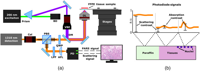
We present a method for direct label-free histopathological assessment of formalin-fixed paraffin-embedded tissue blocks and thin tissue sections using a dual-contrast photoacoustic remote sensing (PARS) microscopy system.

APPROACH

To emulate the nuclear and cellular contrast of H&E staining, we leverage unique properties of the PARS system. Here, the ultraviolet excitation PARS microscope takes advantage of DNA's unique optical absorption to provide nuclear contrast analogous to hematoxylin staining of cell nuclei. Concurrently, the optical scattering contrast of the PARS detection system is leveraged to provide bulk tissue contrast reminiscent of eosin staining of cell membranes.

RESULTS

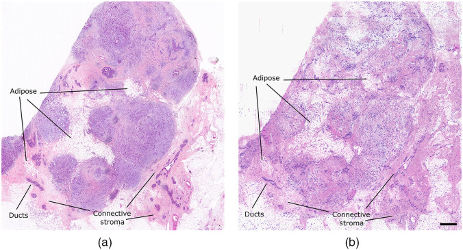
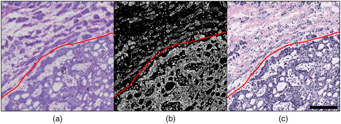
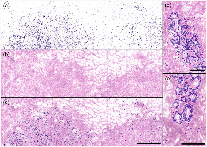
We demonstrate the efficacy of this technique by imaging human breast tissue and human skin tissues in formalin-fixed paraffin-embedded tissue blocks and frozen sections, respectively. Salient nuclear and extranuclear features such as cancerous cells, glands and ducts, adipocytes, and stromal structures such as collagen are captured.

CONCLUSIONS

The presented dual-contrast PARS microscope enables label-free visualization of tissues with contrast and quality comparable to the current gold standard for histopathological analysis. Thus, the proposed system is well positioned to augment existing histopathological workflows, providing histological imaging directly on unstained tissue blocks and sections.

摘要

意义

组织的组织病理学分析是对癌症和其他恶性肿瘤进行分级、分期、诊断和切除的重要工具。目前的组织病理学成像技术需要对样本进行大量处理,然后用苏木精和伊红(H&E)染料进行染色,以突出核和细胞形态。样本制备和染色需要大量的资源,并且在样本制备过程中存在潜在的可变性。

目的

我们提出了一种使用双对比光声远程传感(PARS)显微镜系统对福尔马林固定石蜡包埋组织块和薄组织切片进行直接无标记组织病理学评估的方法。

方法

为了模拟 H&E 染色的核和细胞对比,我们利用 PARS 系统的独特特性。在这里,紫外激发 PARS 显微镜利用 DNA 的独特光吸收特性提供类似于细胞核苏木精染色的核对比。同时,利用 PARS 检测系统的光学散射对比提供类似于细胞膜伊红染色的组织整体对比。

结果

我们分别在福尔马林固定石蜡包埋组织块和冷冻切片中对人乳腺组织和人皮肤组织进行成像,证明了该技术的有效性。捕捉到了明显的核内和核外特征,如癌细胞、腺体和导管、脂肪细胞以及胶原等基质结构。

结论

所提出的双对比 PARS 显微镜能够实现无标记的组织可视化,其对比度和质量可与目前组织病理学分析的金标准相媲美。因此,该系统有望增强现有的组织病理学工作流程,直接在未染色的组织块和切片上提供组织学成像。

https://cdn.ncbi.nlm.nih.gov/pmc/blobs/7b62/8144614/56d6bbf2f8c8/JBO-026-056007-g005.jpg
https://cdn.ncbi.nlm.nih.gov/pmc/blobs/7b62/8144614/34af469d167a/JBO-026-056007-g001.jpg
https://cdn.ncbi.nlm.nih.gov/pmc/blobs/7b62/8144614/250f3575fedb/JBO-026-056007-g002.jpg
https://cdn.ncbi.nlm.nih.gov/pmc/blobs/7b62/8144614/2045f9b08c90/JBO-026-056007-g003.jpg
https://cdn.ncbi.nlm.nih.gov/pmc/blobs/7b62/8144614/132f8ca956c3/JBO-026-056007-g004.jpg
https://cdn.ncbi.nlm.nih.gov/pmc/blobs/7b62/8144614/56d6bbf2f8c8/JBO-026-056007-g005.jpg

相似文献

1
Single acquisition label-free histology-like imaging with dual-contrast photoacoustic remote sensing microscopy.基于双对比光声远程传感显微镜的单采集无标记组织学样成像。
J Biomed Opt. 2021 May;26(5). doi: 10.1117/1.JBO.26.5.056007.
2
Reflection-mode virtual histology using photoacoustic remote sensing microscopy.反射模式虚拟组织学应用于光声远程感测显微镜。
Sci Rep. 2020 Nov 5;10(1):19121. doi: 10.1038/s41598-020-76155-6.
3
Virtual hematoxylin and eosin histopathology using simultaneous photoacoustic remote sensing and scattering microscopy.利用光声远程传感和散射显微镜进行虚拟苏木精和伊红组织病理学检查。
Opt Express. 2021 Apr 26;29(9):13864-13875. doi: 10.1364/OE.423740.
4
Improving maximal safe brain tumor resection with photoacoustic remote sensing microscopy.利用光声遥感显微镜提高最大安全脑肿瘤切除率。
Sci Rep. 2020 Oct 14;10(1):17211. doi: 10.1038/s41598-020-74160-3.
5
F-mode ultraviolet photoacoustic remote sensing for label-free virtual H&E histopathology using a single excitation wavelength.基于单激发波长的 F 模式紫外光声远程传感技术,实现免标记虚拟苏木精-伊红(H&E)组织病理学。
Opt Lett. 2021 Aug 1;46(15):3500-3503. doi: 10.1364/OL.426543.
6
Label-free complete absorption microscopy using second generation photoacoustic remote sensing.基于第二代光声远程传感的无标记全吸收显微镜。
Sci Rep. 2022 May 19;12(1):8464. doi: 10.1038/s41598-022-11235-3.
7
Virtual histological staining of label-free total absorption photoacoustic remote sensing (TA-PARS).无标记全吸收光声远程传感(TA-PARS)的虚拟组织染色。
Sci Rep. 2022 Jun 18;12(1):10296. doi: 10.1038/s41598-022-14042-y.
8
All-optical label-free human breast tissue block histology using photoacoustic remote sensing.基于光声远程传感的全光免标记人体乳腺组织块组织学研究
Opt Lett. 2020 Sep 1;45(17):4770-4773. doi: 10.1364/OL.397223.
9
Virtual histopathology with ultraviolet scattering and photoacoustic remote sensing microscopy.紫外散射和光声远程传感显微镜下的虚拟组织病理学。
Opt Lett. 2021 Oct 15;46(20):5153-5156. doi: 10.1364/OL.436136.
10
Three-dimensional virtual histology in unprocessed resected tissues with photoacoustic remote sensing (PARS) microscopy and optical coherence tomography (OCT).基于光声远程传感(PARS)显微镜和光相干断层扫描(OCT)的未处理切除组织的三维虚拟组织学。
Sci Rep. 2021 Jul 2;11(1):13723. doi: 10.1038/s41598-021-93222-8.

引用本文的文献

1
Metabolic light absorption, scattering, and emission (MetaLASE) microscopy.代谢光吸收、散射和发射(MetaLASE)显微镜。
Sci Adv. 2024 Oct 18;10(42):eadl5729. doi: 10.1126/sciadv.adl5729.
2
Harnessing the power of optical microscopy for visualization and analysis of histopathological images.利用光学显微镜的力量进行组织病理学图像的可视化和分析。
Biomed Opt Express. 2023 Sep 26;14(10):5451-5465. doi: 10.1364/BOE.501893. eCollection 2023 Oct 1.
3
Deep learning-enabled realistic virtual histology with ultraviolet photoacoustic remote sensing microscopy.

本文引用的文献

1
Histopathology for Mohs micrographic surgery with photoacoustic remote sensing microscopy.用于莫氏显微外科手术的组织病理学与光声遥感显微镜技术
Biomed Opt Express. 2020 Dec 24;12(1):654-665. doi: 10.1364/BOE.405869. eCollection 2021 Jan 1.
2
Reflection-mode virtual histology using photoacoustic remote sensing microscopy.反射模式虚拟组织学应用于光声远程感测显微镜。
Sci Rep. 2020 Nov 5;10(1):19121. doi: 10.1038/s41598-020-76155-6.
3
Improving maximal safe brain tumor resection with photoacoustic remote sensing microscopy.利用光声遥感显微镜提高最大安全脑肿瘤切除率。
基于深度学习的紫外光声远程感测显微镜下逼真的虚拟组织学。
Nat Commun. 2023 Sep 25;14(1):5967. doi: 10.1038/s41467-023-41574-2.
4
Three-dimensional virtual histology in unprocessed resected tissues with photoacoustic remote sensing (PARS) microscopy and optical coherence tomography (OCT).基于光声远程传感(PARS)显微镜和光相干断层扫描(OCT)的未处理切除组织的三维虚拟组织学。
Sci Rep. 2021 Jul 2;11(1):13723. doi: 10.1038/s41598-021-93222-8.
Sci Rep. 2020 Oct 14;10(1):17211. doi: 10.1038/s41598-020-74160-3.
4
Serial optical coherence microscopy for label-free volumetric histopathology.用于无标记容积组织病理学的连续光学相干显微镜。
Sci Rep. 2020 Apr 21;10(1):6711. doi: 10.1038/s41598-020-63460-3.
5
Chromophore selective multi-wavelength photoacoustic remote sensing of unstained human tissues.未染色人体组织的发色团选择性多波长光声遥感
Biomed Opt Express. 2019 Oct 1;10(11):5461-5469. doi: 10.1364/BOE.10.005461. eCollection 2019 Nov 1.
6
Ultraviolet photoacoustic remote sensing microscopy.紫外光声遥感显微镜
Opt Lett. 2019 Jul 15;44(14):3586-3589. doi: 10.1364/OL.44.003586.
7
Microscopy with ultraviolet surface excitation for rapid slide-free histology.紫外表面激发显微镜,快速免载玻片组织学。
Nat Biomed Eng. 2017 Dec;1(12):957-966. doi: 10.1038/s41551-017-0165-y. Epub 2017 Dec 4.
8
Non-interferometric photoacoustic remote sensing microscopy.非干涉式光声遥感显微镜
Light Sci Appl. 2017 Jun 2;6(6):e16278. doi: 10.1038/lsa.2016.278. eCollection 2017 Jun.
9
A review of artifacts in histopathology.组织病理学中的伪像综述。
J Oral Maxillofac Pathol. 2018 May-Aug;22(2):279. doi: 10.4103/jomfp.JOMFP_125_15.
10
Light-sheet microscopy for slide-free non-destructive pathology of large clinical specimens.用于大型临床标本的无玻片非破坏性病理学的光片显微镜技术。
Nat Biomed Eng. 2017 Jul;1(7). doi: 10.1038/s41551-017-0084. Epub 2017 Jun 26.