University of Alabama-Birmingham, Birmingham, AL, 35294, USA.
HudsonAlpha Institute for Biotechnology, Huntsville, AL, 35806, USA.
BMC Cancer. 2021 May 29;21(1):632. doi: 10.1186/s12885-021-08388-1.
Pancreatic ductal adenocarcinoma (PDAC) patients suffer poor outcomes, including a five-year survival of below 10%. Poor outcomes result in part from therapeutic resistance that limits the impact of cytotoxic first-line therapy. Novel therapeutic approaches are needed, but currently no targeted therapies exist to treat PDAC.
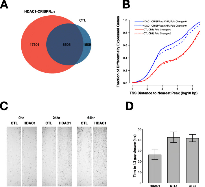
To assess cellular resistance mechanisms common to four cytotoxic chemotherapies (gemcitabine, 5-fluorouracil, irinotecan, and oxaliplatin) used to treat PDAC patients, we performed four genome-wide CRISPR activation (CRISPR) and CRISPR knock-out (CRISPR) screens in two common PDAC cell lines (Panc-1 and BxPC3). We used pathway analysis to identify gene sets enriched among our hits and conducted RNA-sequencing and chromatin immunoprecipitation-sequencing (ChIP-seq) to characterize top hits from our screen. We used scratch assays to assess changes in cellular migration with HDAC1 overexpression.
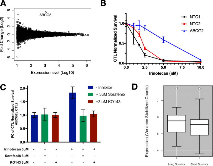
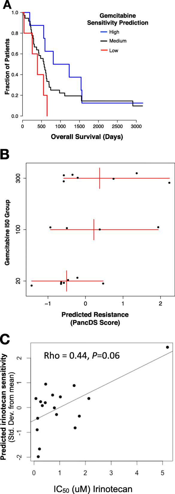
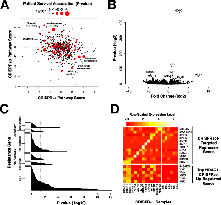
Our data revealed activation of ABCG2, a well-described efflux pump, as the most consistent mediator of resistance in each of our screens. CRISPR-mediated activation of genes involved in transcriptional co-repressor complexes also conferred resistance to multiple drugs. Expression of many of these genes, including HDAC1, is associated with reduced survival in PDAC patients. Up-regulation of HDAC1 in vitro increased promoter occupancy and expression of several genes involved in the epithelial-to-mesenchymal transition (EMT). These cells also displayed phenotypic changes in cellular migration consistent with activation of the EMT pathway. The expression changes resulting from HDAC1 activation were also observed with activation of several other co-repressor complex members. Finally, we developed a publicly available analysis tool, PancDS, which integrates gene expression profiles with our screen results to predict drug sensitivity in resected PDAC tumors and cell lines.
Our results provide a comprehensive resource for identifying cellular mechanisms of drug resistance in PDAC, mechanistically implicate HDAC1, and co-repressor complex members broadly, in multi-drug resistance, and provide an analytical tool for predicting treatment response in PDAC tumors and cell lines.
胰腺导管腺癌 (PDAC) 患者的预后较差,五年生存率低于 10%。治疗效果不佳部分归因于治疗耐药,这限制了细胞毒性一线治疗的效果。需要新的治疗方法,但目前尚无针对 PDAC 的靶向治疗方法。
为了评估四种用于治疗 PDAC 患者的细胞毒性化疗药物(吉西他滨、5-氟尿嘧啶、伊立替康和奥沙利铂)常见的细胞耐药机制,我们在两种常见的 PDAC 细胞系(Panc-1 和 BxPC3)中进行了四次全基因组 CRISPR 激活 (CRISPR) 和 CRISPR 敲除 (CRISPR) 筛选。我们使用通路分析来识别我们的命中富集的基因集,并进行 RNA 测序和染色质免疫沉淀测序 (ChIP-seq) 来描述我们筛选的顶级命中。我们使用划痕实验来评估 HDAC1 过表达对细胞迁移的影响。
我们的数据显示,ABC2,一种众所周知的外排泵,作为我们每次筛选中最一致的耐药介质被激活。涉及转录共抑制复合物的基因的 CRISPR 介导激活也赋予了对多种药物的耐药性。这些基因中的许多基因,包括 HDAC1,的表达与 PDAC 患者的生存时间缩短有关。体外 HDAC1 的上调增加了几个参与上皮间质转化 (EMT) 的基因的启动子占有率和表达。这些细胞在细胞迁移方面也表现出与 EMT 途径激活一致的表型变化。HDAC1 激活导致的表达变化也在其他几个共抑制复合物成员的激活中观察到。最后,我们开发了一个公开的分析工具,PancDS,它将基因表达谱与我们的筛选结果集成在一起,以预测切除的 PDAC 肿瘤和细胞系对药物的敏感性。
我们的研究结果为鉴定 PDAC 中的药物耐药细胞机制提供了一个全面的资源,从机制上广泛涉及 HDAC1 和共抑制复合物成员在多药耐药中的作用,并为预测 PDAC 肿瘤和细胞系的治疗反应提供了一个分析工具。