Ye Qiannan, Zhou Yang, Zhao Changqing, Xu Lieming, Ping Jian
Shuguang Hospital Affiliated to Shanghai University of Traditional Chinese Medicine, Shanghai, China.
Institute of Liver Diseases, Shanghai University of Traditional Chinese Medicine, Shanghai, China.
Front Pharmacol. 2021 May 13;12:677810. doi: 10.3389/fphar.2021.677810. eCollection 2021.
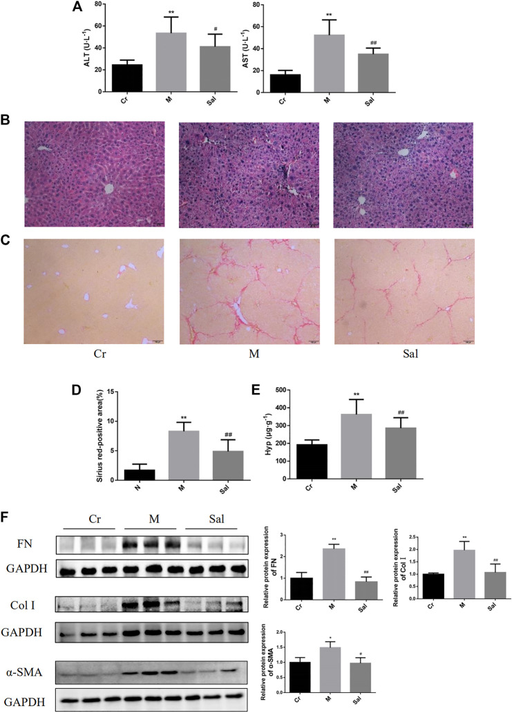
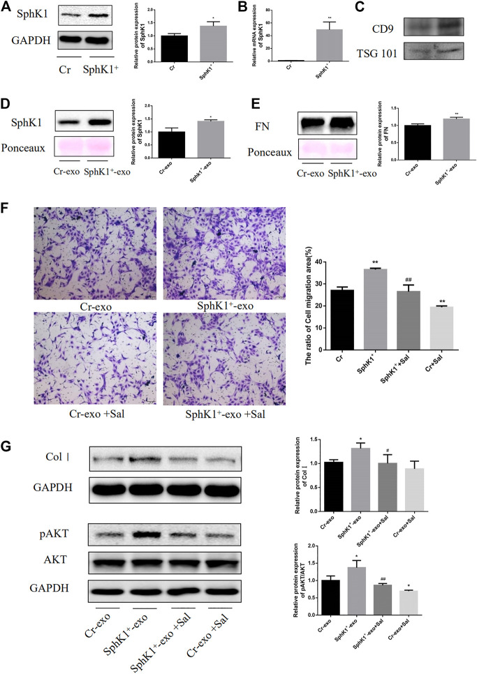
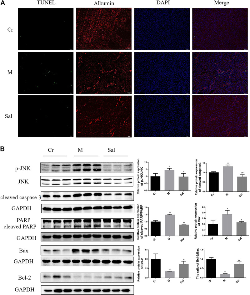
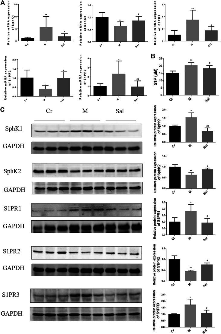
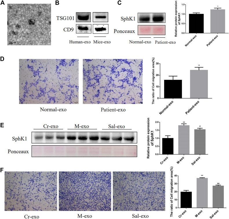
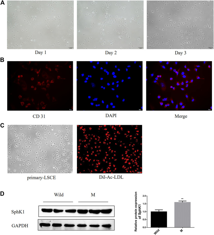
Sphingosine kinase 1 (SphK1)/Sphingosine-1-phosphate (S1P)/S1PRs signaling pathway is known to involve the advancement of liver fibrosis. Exosomal SphK1 promotes hepatic stellate cells (HSC) migration. Salidroside (Sal) inhibits liver fibrosis, but its mechanism is yet to be elucidated. This study was to explore the influences of Sal on the SphK/S1P/S1PRs signaling pathway in liver fibrosis induced by carbon tetrachloride (CCl) , and investigated the mechanism of Sal affecting the migration and activation of HSC triggered by exosomal SphK1 . Our data showed that Sal reduced the activities of alanine transaminase (ALT), aspartate aminotransferase (AST) in serum, and hydroxyproline (Hyp) content in the liver tissue. Sal subdued the expression of α-smooth muscle actin (α-SMA), fibronectin (FN) and type I collagen (Col I) of the liver. Sal also reduced mitochondria-induced hepatocyte apoptosis and to inhibit JNK activation. Furthermore, Sal remarkably eradicated the influence of SphK1, SphK2, S1P, and S1PRs triggered by CCl, whether stimulating or hindering. Compared with serum-derived exosomes from model group mice, serum-derived exosomes from Sal group mice expressed lower SphK1 and reduced JS 1 (mouse HSC cell line) migration. In addition, Sal was also observed to subdue Col I expression, AKT activation, and LX-2 migration induced by exosomal SphK1 from SK-HEP-1 (a kind of liver sinusoidal endothelial cells (LSEC) cell line). In conclusion, Sal could effectively alleviate liver injury, hepatocyte apoptosis, and liver fibrosis , providing supports that the protective effects of Sal might be realized by suppressing JNK activation and modulating the SphK/S1P/S1PRs axis. , it was observed that Sal might alleviate LX-2 migration and activation induced by exosomal SphK1 by inhibiting the AKT activation.
已知鞘氨醇激酶1(SphK1)/1-磷酸鞘氨醇(S1P)/S1P受体(S1PRs)信号通路与肝纤维化进展有关。外泌体SphK1可促进肝星状细胞(HSC)迁移。红景天苷(Sal)可抑制肝纤维化,但其机制尚待阐明。本研究旨在探讨Sal对四氯化碳(CCl)诱导的肝纤维化中SphK/S1P/S1PRs信号通路的影响,并研究Sal影响外泌体SphK1触发的HSC迁移和激活的机制。我们的数据表明,Sal降低了血清中丙氨酸转氨酶(ALT)、天冬氨酸转氨酶(AST)的活性以及肝组织中羟脯氨酸(Hyp)的含量。Sal抑制了肝脏中α平滑肌肌动蛋白(α-SMA)、纤连蛋白(FN)和I型胶原(Col I)的表达。Sal还减少了线粒体诱导的肝细胞凋亡并抑制JNK激活。此外,无论刺激还是抑制,Sal均能显著消除CCl触发的SphK1、SphK2、S1P和S1PRs的影响。与模型组小鼠血清来源的外泌体相比,Sal组小鼠血清来源的外泌体表达较低的SphK1并减少了JS 1(小鼠HSC细胞系)的迁移。此外,还观察到Sal可抑制由SK-HEP-1(一种肝窦内皮细胞(LSEC)细胞系)来源的外泌体SphK1诱导的Col I表达、AKT激活和LX-2迁移。总之,Sal可有效减轻肝损伤、肝细胞凋亡和肝纤维化,这表明Sal的保护作用可能是通过抑制JNK激活和调节SphK/S1P/S1PRs轴来实现的。据观察,Sal可能通过抑制AKT激活来减轻外泌体SphK1诱导的LX-2迁移和激活。