Division of Gastroenterology, Department of Medicine, Haukeland University Hospital, Bergen, Norway.
National Center for Functional Gastrointestinal Disorders, Division of Gastroenterology, Haukeland University Hospital, Bergen, Norway.
Front Cell Infect Microbiol. 2021 May 12;11:524851. doi: 10.3389/fcimb.2021.524851. eCollection 2021.
Interactions between the gut microbiota and enteroendocrine cells play important role in irritable bowel syndrome (IBS). Reduced stem cell densities and their differentiation into enteroendocrine cells may cause abnormal densities of the duodenal enteroendocrine cells in IBS patients.
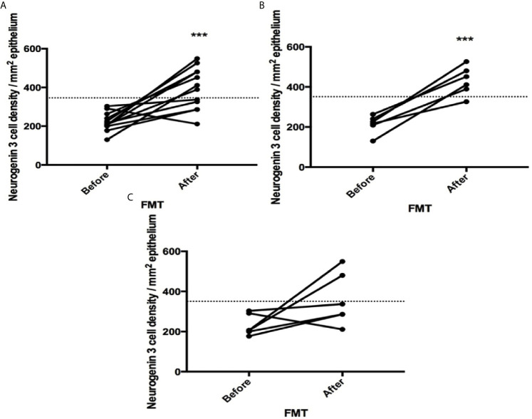
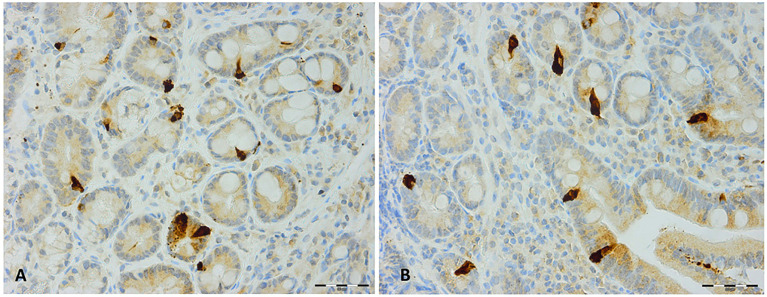
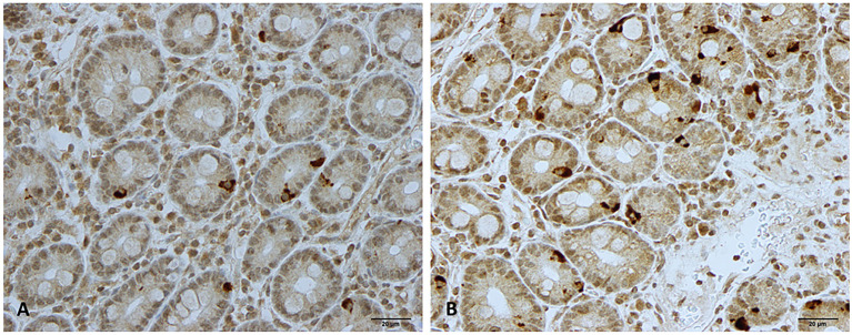
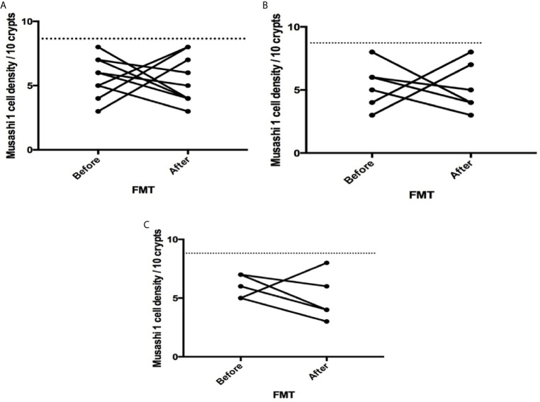
We aimed to investigate the effects of fecal microbiota transplantation (FMT) on stem cell differentiation into enteroendocrine cells as detected by neurogenin 3, stem cells as detected by Musashi 1, and the enteroendocrine cells in the duodenum of IBS patients. The study included 16 IBS patients according to Rome III criteria. Four patients were excluded. The remaining patients (n = 12, four females and eight males) were divided according to the cause of IBS into post-infectious ( = 6) and idiopathic ( = 6) IBS. They completed the following questionnaires before and 3 weeks after FMT: IBS-Symptom Severity Scoring system (IBS-SSS) and IBS-Symptom Questionnaire (IBS-SQ). Feces donated by healthy relatives of the patients were transplanted gastroscope. Biopsies were taken from the descending part of the duodenum at baseline and 3 weeks after FMT. They were immunostained for neurogenin 3, Musashi 1, and all types of duodenal enteroendocrine cells and quantified by computerized image analysis. Microbiota analyses of feces collected just before and 3 weeks after FMT were performed using GA-map™ Dysbiosis test (Genetic Analysis AS, Oslo, Norway).
The total scores for IBS-SSS and IBS-SQ were significantly improved 3 weeks after receiving FMT, = 0.0009 and <0.0001, respectively. The stem cell densities of neurogenin 3 increased significantly following FMT ( = 0.0006) but not for Musashi 1 ( = 0.42). The cell densities of chromogranin A, cholecystokinin, gastric inhibitory peptide, serotonin, and somatostatin, but not for secretin, have significantly changed in both IBS groups after 3 weeks from receiving FMT.
More than two-thirds of IBS patients experienced improvement in their symptoms parallel to changes in the enteroendocrine cells densities 3 weeks after FMT. The changes in the enteroendocrine cell densities do not appear to be caused by changes in the stem cells or their early progenitors rather by changes in the differentiation progeny as detected by neurogenin 3. The study was retrospectively registered at ClinicalTrials.gov (ID: NCT03333291).
ClinicalTrials.gov, identifier NCT03333291.
肠道微生物群与肠内分泌细胞的相互作用在肠易激综合征(IBS)中起着重要作用。干细胞密度的降低及其向肠内分泌细胞的分化可能导致 IBS 患者十二指肠肠内分泌细胞的异常密度。
我们旨在研究粪便微生物群移植(FMT)对神经基因 3 检测的干细胞分化为肠内分泌细胞、Musashi 1 检测的干细胞以及 IBS 患者十二指肠肠内分泌细胞的影响。该研究纳入了 16 名符合罗马 III 标准的 IBS 患者。其中 4 名患者被排除。其余患者(n=12,4 名女性和 8 名男性)根据 IBS 的病因分为感染后(n=6)和特发性(n=6)IBS。他们在 FMT 前和 3 周后完成了 IBS-症状严重程度评分系统(IBS-SSS)和 IBS-症状问卷(IBS-SQ)。通过胃镜将患者亲属捐献的粪便移植到患者体内。在基线和 FMT 后 3 周时,从十二指肠降部采集活检标本。它们通过免疫组织化学染色用于神经基因 3、Musashi 1 和所有类型的十二指肠肠内分泌细胞,并通过计算机图像分析进行定量。使用 GA-map™ 失调测试(挪威奥斯陆遗传分析公司)在 FMT 前后收集的粪便进行微生物组分析。
接受 FMT 后 3 周,IBS-SSS 和 IBS-SQ 的总评分显著改善,分别为 = 0.0009 和 <0.0001。神经基因 3 的干细胞密度在 FMT 后显著增加(=0.0006),但 Musashi 1 没有增加(=0.42)。在接受 FMT 3 周后,两组 IBS 患者的嗜铬粒蛋白 A、胆囊收缩素、胃抑制肽、血清素和生长抑素的细胞密度均发生显著变化,但分泌素没有变化。
超过三分之二的 IBS 患者在接受 FMT 后 3 周内症状得到改善,同时肠内分泌细胞密度也发生改变。肠内分泌细胞密度的变化似乎不是由干细胞或其早期祖细胞的变化引起的,而是由神经基因 3 检测到的分化后代的变化引起的。该研究在 ClinicalTrials.gov(ID:NCT03333291)进行了回顾性注册。
ClinicalTrials.gov,标识符 NCT03333291。