Kihira Shingo, Tsankova Nadejda M, Bauer Adam, Sakai Yu, Mahmoudi Keon, Zubizarreta Nicole, Houldsworth Jane, Khan Fahad, Salamon Noriko, Hormigo Adilia, Nael Kambiz
Department of Diagnostic, Molecular and Interventional Radiology, Icahn School of Medicine at Mount Sinai, New York, New York, USA.
Department of Pathology, Icahn School of Medicine at Mount Sinai, New York, New York, USA.
Neurooncol Adv. 2021 Apr 8;3(1):vdab051. doi: 10.1093/noajnl/vdab051. eCollection 2021 Jan-Dec.
Early identification of glioma molecular phenotypes can lead to understanding of patient prognosis and treatment guidance. We aimed to develop a multiparametric MRI texture analysis model using a combination of conventional and diffusion MRI to predict a wide range of biomarkers in patients with glioma.
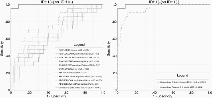
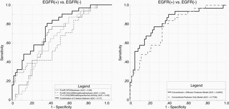
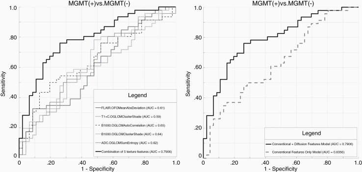
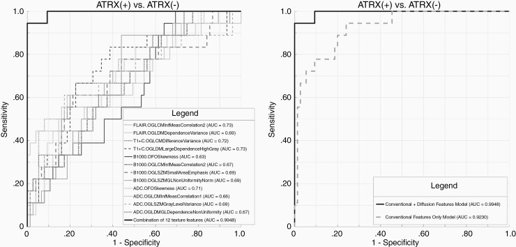
In this retrospective study, patients were included if they (1) had diagnosis of gliomas with known , , , , , and status from surgical pathology and (2) had preoperative MRI including FLAIR, T1c+ and diffusion for radiomic texture analysis. Statistical analysis included logistic regression and receiver-operating characteristic (ROC) curve analysis to determine the optimal model for predicting glioma biomarkers. A comparative analysis between ROCs (conventional only vs conventional + diffusion) was performed.
From a total of 111 patients included, 91 (82%) were categorized to training and 20 (18%) to test datasets. Constructed cross-validated model using a combination of texture features from conventional and diffusion MRI resulted in overall AUC/accuracy of 1/79% for , 0.99/80% for , 0.79/67% for , and 0.77/66% for . The addition of diffusion data to conventional MRI features significantly ( < .05) increased predictive performance for , , and . The overall accuracy of the final model in predicting biomarkers in the test group was 80% (), 70% (), 70% (), and 75% ().
Addition of MR diffusion to conventional MRI features provides added diagnostic value in preoperative determination of IDH1, MGMT, and ATRX in patients with glioma.
早期识别胶质瘤分子表型有助于了解患者预后并指导治疗。我们旨在开发一种多参数MRI纹理分析模型,结合传统MRI和弥散MRI来预测胶质瘤患者的多种生物标志物。
在这项回顾性研究中,纳入的患者需满足以下条件:(1)经手术病理确诊为胶质瘤,且已知其 、 、 、 、 和 状态;(2)术前行MRI检查,包括FLAIR、T1c +和弥散加权成像,用于进行影像组学纹理分析。统计分析包括逻辑回归和受试者工作特征(ROC)曲线分析,以确定预测胶质瘤生物标志物的最佳模型。对ROC(仅传统MRI与传统MRI +弥散MRI)进行了比较分析。
总共纳入111例患者,其中91例(82%)被分类到训练数据集,20例(18%)被分类到测试数据集。使用传统MRI和弥散MRI的纹理特征组合构建的交叉验证模型,对于 ,总体AUC/准确率为1/79%;对于 ,为0.99/80%;对于 ,为0.79/67%;对于 ,为0.77/66%。将弥散数据添加到传统MRI特征中,显著( <.05)提高了对 、 和 的预测性能。最终模型在测试组中预测生物标志物的总体准确率分别为80%()、70%()、70%()和75%()。
在传统MRI特征中加入磁共振弥散成像,在术前确定胶质瘤患者的IDH1、MGMT和ATRX时具有额外的诊断价值。