Institute of Medical Biology, Chinese Academy of Medical Sciences and Peking Union Medical College, Kunming, People's Republic of China.
Emerg Microbes Infect. 2021 Dec;10(1):1156-1168. doi: 10.1080/22221751.2021.1938241.
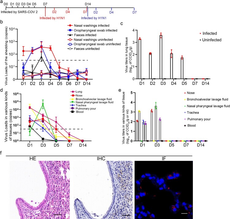
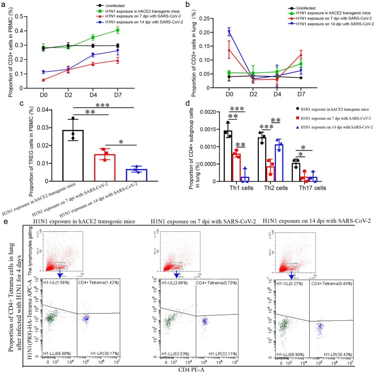
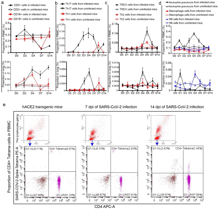
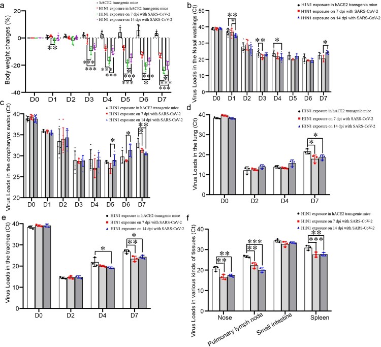
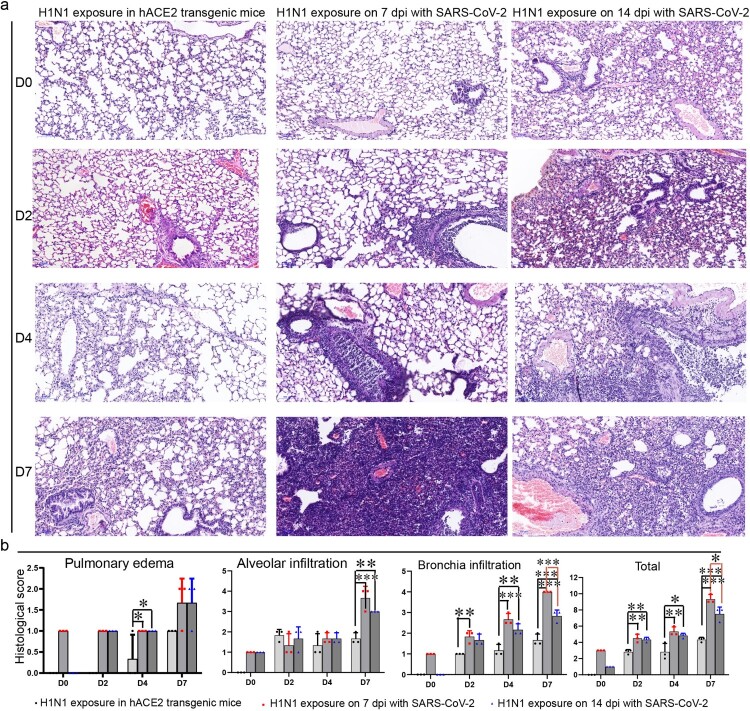
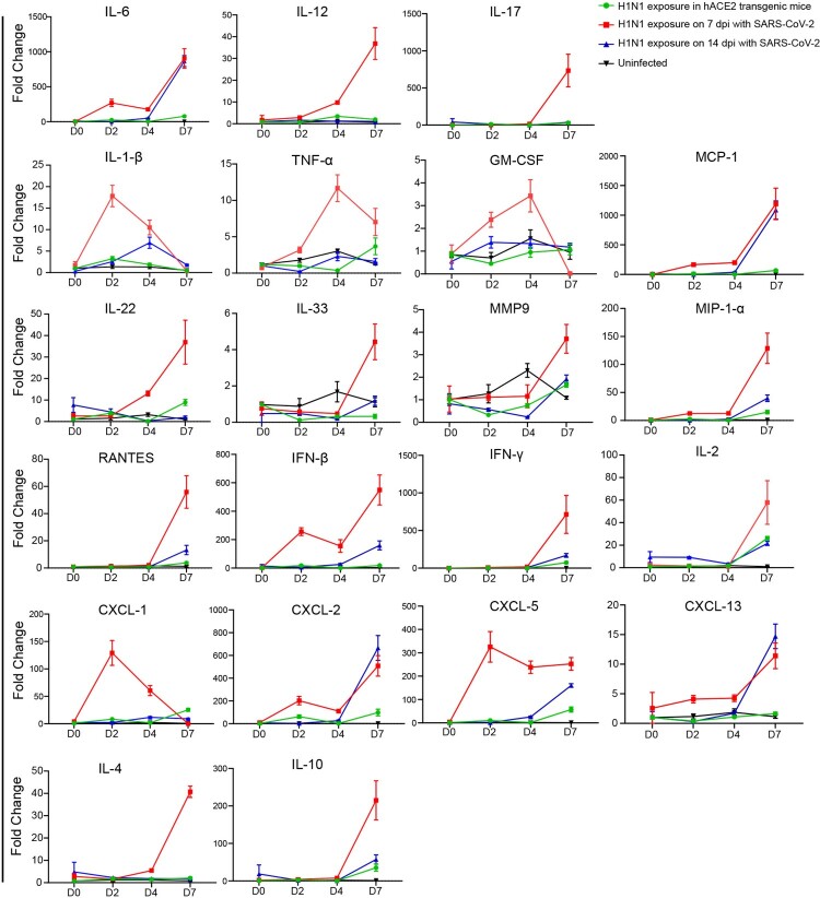
The risk of secondary infection with SARS-CoV-2 and influenza A virus is becoming a practical problem that must be addressed as the flu season merges with the COVID-19 pandemic. As SARS-CoV-2 and influenza A virus have been found in patients, understanding the in vivo characteristics of the secondary infection between these two viruses is a high priority. Here, hACE2 transgenic mice were challenged with the H1N1 virus at a nonlethal dose during the convalescent stage on 7 and 14 days post SARS-CoV-2 infection, and importantly, subsequent H1N1 infection showed enhanced viral shedding and virus tissue distribution. Histopathological observation revealed an extensive pathological change in the lungs related to H1N1 infection in mice recovered from SARS-CoV-2 infection, with severe inflammation infiltration and bronchiole disruption. Moreover, upon H1N1 exposure on 7 and 14 dpi of SARS-CoV-2 infection, the lymphocyte population activated at a lower level with T cell suppressed in both PBMC and lung. These findings will be valuable for evaluating antiviral therapeutics and vaccines as well as guiding public health work.
SARS-CoV-2 和甲型流感病毒(influenza A virus)二次感染的风险正在成为一个现实问题,因为流感季节与 COVID-19 大流行同时出现。由于已经在患者体内发现了 SARS-CoV-2 和甲型流感病毒,因此了解这两种病毒之间二次感染的体内特征是当务之急。在这里,在感染 SARS-CoV-2 后第 7 天和第 14 天的恢复期,用非致死剂量的 H1N1 病毒对 hACE2 转基因小鼠进行了挑战,重要的是,随后的 H1N1 感染显示出病毒脱落和病毒组织分布增加。组织病理学观察显示,从 SARS-CoV-2 感染中恢复的小鼠肺部与 H1N1 感染相关的广泛病理变化,严重的炎症浸润和细支气管破坏。此外,在 SARS-CoV-2 感染后第 7 天和第 14 天接触 H1N1 时,在 PBMC 和肺部中 T 细胞受到抑制的情况下,淋巴细胞群体的激活水平较低。这些发现对于评估抗病毒治疗药物和疫苗以及指导公共卫生工作将是有价值的。