Bojin Florina, Robu Andreea, Bejenariu Maria Iulia, Ordodi Valentin, Olteanu Emilian, Cean Ada, Popescu Roxana, Neagu Monica, Gavriliuc Oana, Neagu Adrian, Arjoca Stelian, Păunescu Virgil
Department of Functional Sciences, Victor Babes University of Medicine and Pharmacy Timisoara, 300041 Timisoara, Romania.
OncoGen Institute, 300723 Timisoara, Romania.
Micromachines (Basel). 2021 May 9;12(5):535. doi: 10.3390/mi12050535.
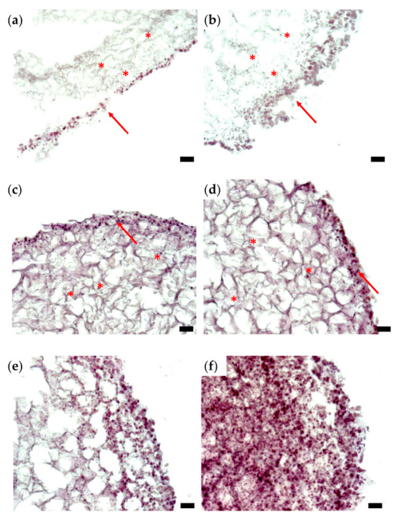
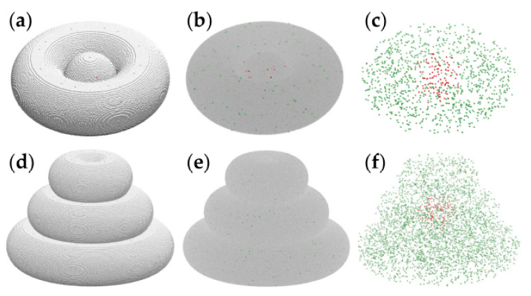
The tumor microenvironment (TME) influences cancer progression. Therefore, engineered TME models are being developed for fundamental research and anti-cancer drug screening. This paper reports the biofabrication of 3D-printed avascular structures that recapitulate several features of the TME. The tumor is represented by a hydrogel droplet uniformly loaded with breast cancer cells (10 cells/mL); it is embedded in the same type of hydrogel containing primary cells-tumor-associated fibroblasts isolated from the peritumoral environment and peripheral blood mononuclear cells. Hoechst staining of cryosectioned tissue constructs demonstrated that cells remodeled the hydrogel and remained viable for weeks. Histological sections revealed heterotypic aggregates of malignant and peritumoral cells; moreover, the constituent cells proliferated in vitro. To investigate the interactions responsible for the experimentally observed cellular rearrangements, we built lattice models of the bioprinted constructs and simulated their evolution using Metropolis Monte Carlo methods. Although unable to replicate the complexity of the TME, the approach presented here enables the self-assembly and co-culture of several cell types of the TME. Further studies will evaluate whether the bioprinted constructs can evolve in vivo in animal models. If they become connected to the host vasculature, they may turn into a fully organized TME.
肿瘤微环境(TME)影响癌症进展。因此,正在开发工程化的TME模型用于基础研究和抗癌药物筛选。本文报道了3D打印无血管结构的生物制造,该结构概括了TME的几个特征。肿瘤由均匀负载乳腺癌细胞(10个细胞/毫升)的水凝胶微滴代表;它被嵌入含有从肿瘤周围环境分离的原代细胞——肿瘤相关成纤维细胞和外周血单核细胞的同类型水凝胶中。对冷冻切片组织构建体进行的Hoechst染色表明,细胞重塑了水凝胶并存活了数周。组织学切片显示恶性细胞和肿瘤周围细胞的异型聚集;此外,组成细胞在体外增殖。为了研究导致实验观察到的细胞重排的相互作用,我们构建了生物打印构建体的晶格模型,并使用 metropolis 蒙特卡罗方法模拟它们的演化。尽管无法复制TME的复杂性,但本文提出的方法能够实现TME几种细胞类型的自组装和共培养。进一步的研究将评估生物打印构建体是否能在动物模型体内演化。如果它们与宿主脉管系统相连,它们可能会变成一个完全组织化的TME。