• 文献检索
  • 文档翻译
  • 深度研究
  • 学术资讯
  • Suppr Zotero 插件Zotero 插件
  • 邀请有礼
  • 套餐&价格
  • 历史记录
应用&插件
Suppr Zotero 插件Zotero 插件浏览器插件Mac 客户端Windows 客户端微信小程序
定价
高级版会员购买积分包购买API积分包
服务
文献检索文档翻译深度研究API 文档MCP 服务
关于我们
关于 Suppr公司介绍联系我们用户协议隐私条款
关注我们

Suppr 超能文献

核心技术专利:CN118964589B侵权必究
粤ICP备2023148730 号-1Suppr @ 2026

文献检索

告别复杂PubMed语法,用中文像聊天一样搜索,搜遍4000万医学文献。AI智能推荐,让科研检索更轻松。

立即免费搜索

文件翻译

保留排版,准确专业,支持PDF/Word/PPT等文件格式,支持 12+语言互译。

免费翻译文档

深度研究

AI帮你快速写综述,25分钟生成高质量综述,智能提取关键信息,辅助科研写作。

立即免费体验

基于叠加的头影测量法定量颅面变化。

A Superimposition-Based Cephalometric Method to Quantitate Craniofacial Changes.

机构信息

Orthodontics, Department of Odontology, Umeå University, SE-901 85 Umeå, Sweden.

Oral and Maxillofacial Radiology, Department of Odontology, Umeå University, SE-901 85 Umeå, Sweden.

出版信息

Int J Environ Res Public Health. 2021 May 14;18(10):5260. doi: 10.3390/ijerph18105260.

DOI:10.3390/ijerph18105260
PMID:34069290
原文链接:https://pmc.ncbi.nlm.nih.gov/articles/PMC8156959/
Abstract

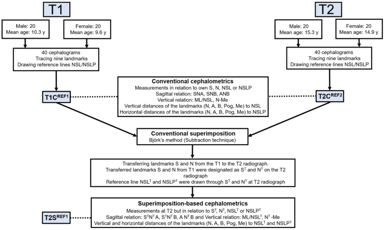
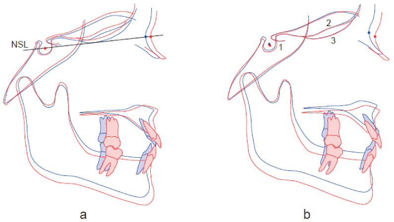
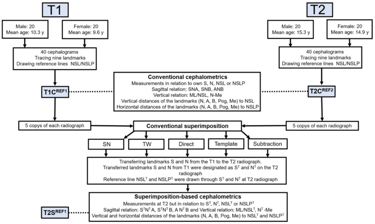
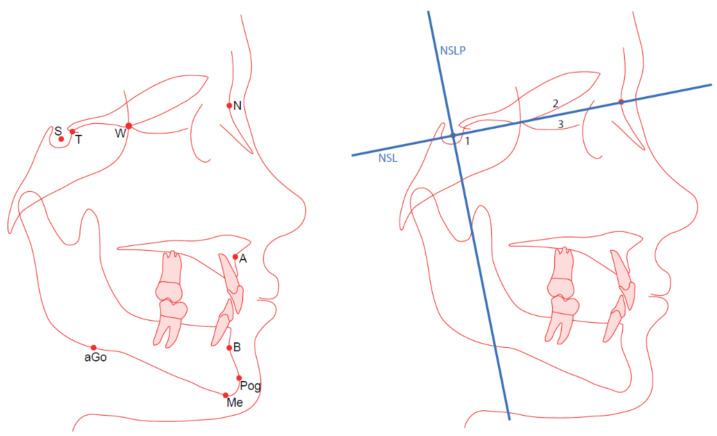
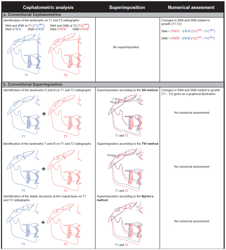
To assess the craniofacial changes related to growth and/or to orthodontic and orthognathic treatments, it is necessary to superimpose serial radiographs on stable structures. However, conventional superimposition provides only a graphical illustration of these changes. To increase the precision of growth and treatment evaluations, it is desirable to quantitate these craniofacial changes. The aims of this study were to (1) evaluate a superimposition-based cephalometric method to process numerical data for craniofacial growth changes and (2) identify a valid, reliable, and feasible method for superimposition. Forty pairs of cephalograms were analyzed at T1 and T2 (mean age 9.9 and 15.0 years, respectively). The superimposition-based cephalometric method involved relating the sagittal and vertical measurements on the T2 radiographs to the nasion and sella landmarks on the T1 radiographs. Validity and reliability were evaluated for three superimposition methods: the sella-nasion (SN); the tuberculum sella-wing (TW); and Björk's structural. Superimposition-based cephalometrics can be used to quantify craniofacial changes digitally. The numerical data from the superimposition-based cephalometrics reflected a graphical illustration of superimposition and differed significantly from the data acquired through conventional cephalometrics. Superimposition using the TW method is recommended as it is valid, reliable, and feasible.

摘要

为了评估与生长和/或正畸和正颌治疗相关的颅面变化,有必要将连续的射线照片叠加到稳定的结构上。然而,传统的叠加仅提供了这些变化的图形说明。为了提高生长和治疗评估的精度,量化这些颅面变化是可取的。本研究的目的是:(1)评估一种基于叠加的头影测量方法来处理颅面生长变化的数值数据;(2)确定一种有效、可靠和可行的叠加方法。在 T1 和 T2 时分析了 40 对头颅侧位片(平均年龄分别为 9.9 岁和 15.0 岁)。基于叠加的头影测量方法涉及将 T2 射线照片上的矢状和垂直测量值与 T1 射线照片上的鼻根和蝶鞍标志点相关联。评估了三种叠加方法的有效性和可靠性:蝶鞍-鼻根(SN);蝶鞍-翼突(TW);和 Björk 的结构。基于叠加的头影测量法可用于数字化定量颅面变化。基于叠加的头影测量法的数值数据反映了叠加的图形说明,与通过传统头影测量法获得的数据有显著差异。建议使用 TW 方法进行叠加,因为它是有效的、可靠的和可行的。

https://cdn.ncbi.nlm.nih.gov/pmc/blobs/f405/8156959/d03c80b6b49a/ijerph-18-05260-g004.jpg
https://cdn.ncbi.nlm.nih.gov/pmc/blobs/f405/8156959/ce32bfe022a1/ijerph-18-05260-g0A1.jpg
https://cdn.ncbi.nlm.nih.gov/pmc/blobs/f405/8156959/dd2c324fb5a7/ijerph-18-05260-g0A2.jpg
https://cdn.ncbi.nlm.nih.gov/pmc/blobs/f405/8156959/7c156f4f3902/ijerph-18-05260-g001.jpg
https://cdn.ncbi.nlm.nih.gov/pmc/blobs/f405/8156959/9c4fd771d34f/ijerph-18-05260-g002a.jpg
https://cdn.ncbi.nlm.nih.gov/pmc/blobs/f405/8156959/ef63615861cf/ijerph-18-05260-g003.jpg
https://cdn.ncbi.nlm.nih.gov/pmc/blobs/f405/8156959/d03c80b6b49a/ijerph-18-05260-g004.jpg

相似文献

1
A Superimposition-Based Cephalometric Method to Quantitate Craniofacial Changes.基于叠加的头影测量法定量颅面变化。
Int J Environ Res Public Health. 2021 May 14;18(10):5260. doi: 10.3390/ijerph18105260.
2
Evaluation of an automated superimposition method based on multiple landmarks for growing patients.基于多个标志点的生长患者自动配准方法的评估。
Angle Orthod. 2022 Mar 1;92(2):226-232. doi: 10.2319/010121-1.1.
3
Longitudinal growth changes of the cranial base from puberty to adulthood. A comparison of different superimposition methods.颅基底从青春期到成年的纵向生长变化。不同叠加方法的比较。
Angle Orthod. 2010 Jul;80(4):537-44. doi: 10.2319/080709-447.1.
4
Evaluation of an automated superimposition method for computer-aided cephalometrics.自动叠加法在计算机辅助头影测量中的评价。
Angle Orthod. 2020 May 1;90(3):390-396. doi: 10.2319/071319-469.1.
5
Radiographic evaluation of orthodontic treatment by means of four different cephalometric superimposition methods.通过四种不同的头影测量重叠方法对正畸治疗进行影像学评估。
Dental Press J Orthod. 2015 May-Jun;20(3):29-36. doi: 10.1590/2176-9451.20.3.029-036.oar.
6
Cephalometric superimposition on the occipital condyles as a longitudinal growth assessment reference: I-point and I-curve.以枕髁为纵向生长评估参考的头影测量重叠法:I点和I曲线。
Anat Rec (Hoboken). 2008 Dec;291(12):1603-10. doi: 10.1002/ar.20761.
7
Assessing lower incisor inclination change: a comparison of four cephalometric methods.评估下切牙倾斜度变化:四种头影测量方法的比较
Eur J Orthod. 2016 Apr;38(2):184-9. doi: 10.1093/ejo/cjv027. Epub 2015 Apr 17.
8
The accuracy of cephalometric tracing superimposition.头影测量描记重叠法的准确性。
J Oral Maxillofac Surg. 2006 Feb;64(2):194-202. doi: 10.1016/j.joms.2005.10.028.
9
The validity of two methods of mandibular superimposition: a comparison with tantalum implants.两种下颌骨叠加方法的有效性:与钽植入物的比较
Am J Orthod Dentofacial Orthop. 1998 Mar;113(3):263-70. doi: 10.1016/s0889-5406(98)70295-1.
10
Evaluation of extraction and non-extraction treatment effects by two different superimposition methods.两种不同叠加方法评价拔牙和非拔牙治疗效果。
Eur J Orthod. 2011 Dec;33(6):691-9. doi: 10.1093/ejo/cjq143. Epub 2011 Mar 4.

引用本文的文献

1
Evaluation of an Automatic Cephalometric Superimposition Method Based on Feature Matching.基于特征匹配的自动头影测量叠加方法的评估
J Imaging Inform Med. 2025 Feb 25. doi: 10.1007/s10278-025-01447-0.
2
Skeletal stability after maxillary distraction osteogenesis or conventional Le Fort I osteotomy in patients with cleft lip and palate: A superimposition-based cephalometric analysis.腭裂患者上颌骨牵引成骨或传统 Le Fort I 截骨术后的骨骼稳定性:基于叠加的头影测量分析。
Oral Maxillofac Surg. 2024 Jun;28(2):925-934. doi: 10.1007/s10006-024-01227-0. Epub 2024 Feb 16.
3
Comparison of Diagnostic Validity of Cephalometric Analyses of the ANB Angle and Tau Angle for Assessment of the Sagittal Relationship of Jaw and Mandible.

本文引用的文献

1
Evaluation of pulp cavity/chamber changes after tooth-borne and bone-borne rapid maxillary expansions: a CBCT study using surface-based superimposition and deviation analysis.基于表面叠加和偏差分析的 CBCT 研究评价牙源性和骨源性上颌快速扩弓后牙髓腔/室的变化。
Clin Oral Investig. 2021 Apr;25(4):2237-2247. doi: 10.1007/s00784-020-03539-3. Epub 2020 Aug 29.
2
One Step before 3D Printing-Evaluation of Imaging Software Accuracy for 3-Dimensional Analysis of the Mandible: A Comparative Study Using a Surface-to-Surface Matching Technique.3D打印前的一步——下颌骨三维分析成像软件准确性评估:一项使用表面到表面匹配技术的比较研究
Materials (Basel). 2020 Jun 21;13(12):2798. doi: 10.3390/ma13122798.
3
用于评估颌骨和下颌骨矢状关系的ANB角和Tau角头影测量分析的诊断有效性比较。
J Clin Med. 2023 Oct 2;12(19):6333. doi: 10.3390/jcm12196333.
4
Dentoskeletal and soft tissue changes after treatment of crowding with premolar extractions: a 50-year follow-up.拔牙矫治牙列拥挤:50 年的随访研究牙颌骨及软组织变化。
Eur J Orthod. 2023 Feb 10;45(1):79-87. doi: 10.1093/ejo/cjac035.
5
Craniofacial changes from 13 to 62 years of age.从 13 岁到 62 岁的颅面变化。
Eur J Orthod. 2022 Sep 19;44(5):556-565. doi: 10.1093/ejo/cjac011.
6
Three-Dimensional Analysis of Posterior Mandibular Displacement in Rats.大鼠下颌骨后移位的三维分析
Vet Sci. 2022 Mar 20;9(3):144. doi: 10.3390/vetsci9030144.
7
Early extractions of premolars reduce age-related crowding of lower incisors: 50 years of follow-up.早期拔除前磨牙可减少下切牙与年龄相关的拥挤:50 年的随访研究。
Clin Oral Investig. 2022 Jun;26(6):4525-4535. doi: 10.1007/s00784-022-04416-x. Epub 2022 Feb 24.
Clinical indications and radiation doses of cone beam computed tomography in orthodontics.
口腔正畸锥形束计算机断层扫描的临床适应证及辐射剂量
Med Pharm Rep. 2019 Oct;92(4):346-351. doi: 10.15386/mpr-1434. Epub 2019 Oct 25.
4
Voxel-based superimposition of serial craniofacial CBCTs: Reliability, reproducibility and segmentation effect on hard-tissue outcomes.基于体素的颅颌面锥形束 CT 序列叠加:可靠性、可重复性和对硬组织结果的分割效果。
Orthod Craniofac Res. 2020 Feb;23(1):92-101. doi: 10.1111/ocr.12347. Epub 2019 Oct 1.
5
Precision and reliability of Dolphin 3-dimensional voxel-based superimposition.Dolphin基于体素的三维叠加的精度和可靠性。
Am J Orthod Dentofacial Orthop. 2018 Apr;153(4):599-606. doi: 10.1016/j.ajodo.2017.07.025.
6
Radiation dose of cone-beam computed tomography compared to conventional radiographs in orthodontics.正畸治疗中锥束计算机断层扫描与传统X线片的辐射剂量比较
J Orofac Orthop. 2016 Jan;77(1):9-15. doi: 10.1007/s00056-015-0002-4. Epub 2016 Jan 8.
7
Radiographic evaluation of orthodontic treatment by means of four different cephalometric superimposition methods.通过四种不同的头影测量重叠方法对正畸治疗进行影像学评估。
Dental Press J Orthod. 2015 May-Jun;20(3):29-36. doi: 10.1590/2176-9451.20.3.029-036.oar.
8
Anterior cranial-base time-related changes: A systematic review.前颅底的时间相关变化:一项系统评价。
Am J Orthod Dentofacial Orthop. 2014 Jul;146(1):21-32.e6. doi: 10.1016/j.ajodo.2014.03.019.
9
Comparison of adult and child radiation equivalent doses from 2 dental cone-beam computed tomography units.比较两种口腔锥形束 CT 设备的成人和儿童辐射当量剂量。
Am J Orthod Dentofacial Orthop. 2013 Jun;143(6):784-92. doi: 10.1016/j.ajodo.2013.01.013.
10
Longitudinal growth changes of the cranial base from puberty to adulthood. A comparison of different superimposition methods.颅基底从青春期到成年的纵向生长变化。不同叠加方法的比较。
Angle Orthod. 2010 Jul;80(4):537-44. doi: 10.2319/080709-447.1.