Guijarro Luis G, Sanmartin-Salinas Patricia, Pérez-Cuevas Eva, Toledo-Lobo M Val, Monserrat Jorge, Zoullas Sofía, Sáez Miguel A, Álvarez-Mon Miguel A, Bujan Julia, Noguerales-Fraguas Fernando, Arilla-Ferreiro Eduardo, Álvarez-Mon Melchor, Ortega Miguel A
Unit of Biochemistry and Molecular Biology (CIBEREHD), Department of System Biology, University of Alcalá, 28801 Alcala de Henares, Spain.
Ramón y Cajal Institute of Sanitary Research (IRYCIS), 28034 Madrid, Spain.
Cancers (Basel). 2021 May 23;13(11):2560. doi: 10.3390/cancers13112560.
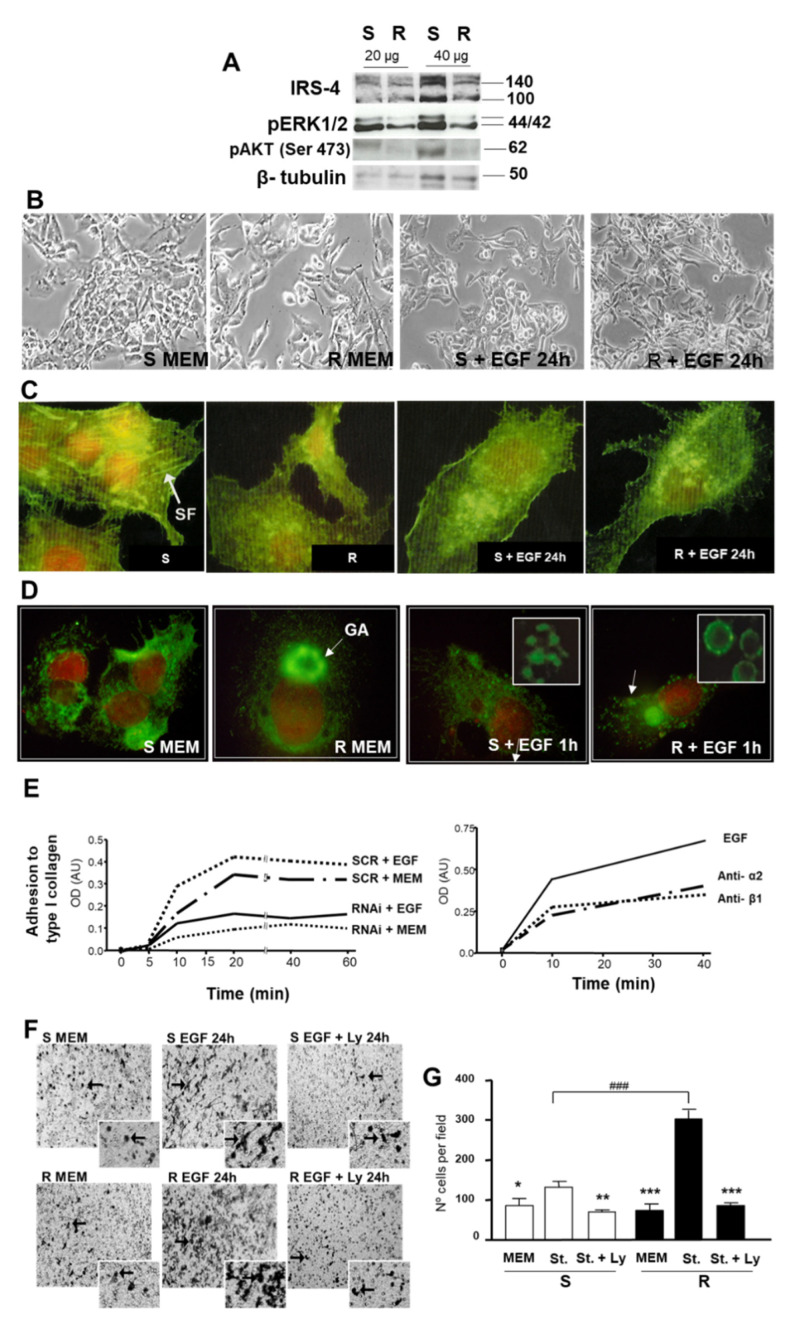
New evidence suggests that insulin receptor substrate 4 (IRS-4) may play an important role in the promotion of tumoral growth. In this investigation, we have evaluated the role of IRS-4 in a pilot study performed on patients with liver cancer. We used immunohistochemistry to examine IRS-4 expression in biopsies of tumoral tissue from a cohort of 31 patient suffering of hepatocellular carcinoma (HCC). We simultaneously analyzed the expression of the cancer biomarkers PCNA, Ki-67, and pH3 in the same tissue samples. The in vitro analysis was conducted by studying the behavior of HepG2 cells following IRS-4 overexpression/silencing. IRS-4 was expressed mainly in the nuclei of tumoral cells from HCC patients. In contrast, in healthy cells involved in portal triads, canaliculi, and parenchymal tissue, IRS-4 was observed in the cytosol and the membrane. Nuclear IRS-4 in the tumoral region was found in 69.9 ± 3.2%, whereas in the surrounding healthy hepatocytes, nuclear IRS-4 was rarely observed. The percentage of tumoral cells that exhibited nuclear PCNA and Ki-67 were 52.1 ± 7%, 6.1 ± 1.1% and 1.3 ± 0.2%, respectively. Furthermore, we observed a significant positive linear correlation between nuclear IRS-4 and PCNA (r = 0.989; < 0.001). However, when we correlated the nuclear expression of IRS-4 and Ki-67, we observed a significant positive curvilinear correlation (r = 0.758; < 0.010). This allowed us to define two populations, (IRS-4 + Ki-67 ≤ 69%) and (IRS-4 + Ki-67 > 70%). The population with lower levels of IRS-4 and Ki-67 had a higher risk of suffering from multifocal liver cancer (OR = 16.66; CI = 1.68-164.8 (95%); < 0.05). Immunoblot analyses showed that IRS-4 in normal human liver biopsies was lower than in HepG2, Huh7, and Chang cells. Treatment of HepG2 with IGF-1 and EGF induced IRS-4 translocation to the nucleus. Regulation of IRS-4 levels via HepG2 transfection experiments revealed the protein's role in proliferation, cell migration, and cell-. Nuclear IRS-4 is increased in the tumoral region of HCC. IRS-4 and Ki-67 levels are significantly correlated with the presence of multifocal HCC. Moreover, upregulation of IRS-4 in HepG2 cells induced proliferation by a β-catenin/Rb/cyclin D mechanism, whereas downregulation of IRS-4 caused a loss in cellular polarity and in its adherence to collagen as well as a gain in migratory and invasive capacities, probably via an integrin α2 and focal adhesion cascade (FAK) mechanism.
新证据表明,胰岛素受体底物4(IRS - 4)可能在促进肿瘤生长中发挥重要作用。在本研究中,我们在一项针对肝癌患者的初步研究中评估了IRS - 4的作用。我们使用免疫组织化学方法检测了31例肝细胞癌(HCC)患者肿瘤组织活检中IRS - 4的表达。我们同时分析了同一组织样本中癌症生物标志物PCNA、Ki - 67和pH3的表达。体外分析通过研究IRS - 4过表达/沉默后HepG2细胞的行为来进行。IRS - 4主要在HCC患者肿瘤细胞的细胞核中表达。相比之下,在门静脉三联管、胆小管和实质组织中的健康细胞中,IRS - 4在细胞质和细胞膜中被观察到。肿瘤区域细胞核中IRS - 4的比例为69.9±3.2%,而在周围健康肝细胞中,很少观察到细胞核IRS - 4。显示细胞核PCNA和Ki - 67的肿瘤细胞百分比分别为52.1±7%、6.1±1.1%和1.3±0.2%。此外,我们观察到细胞核IRS - 4与PCNA之间存在显著的正线性相关性(r = 0.989;<0.001)。然而,当我们将IRS - 4的核表达与Ki - 67进行相关性分析时,我们观察到显著的正曲线相关性(r = 0.758;<0.010)。这使我们能够定义两个群体,(IRS - 4 + Ki - 67≤69%)和(IRS - 4 + Ki - 67>70%)。IRS - 4和Ki - 67水平较低的群体患多灶性肝癌的风险更高(OR = 16.66;CI = 1.68 - 164.8(95%);<0.05)。免疫印迹分析表明,正常人肝活检中的IRS - 4低于HepG2、Huh7和Chang细胞。用IGF - 1和EGF处理HepG2诱导IRS - 4转位至细胞核。通过HepG2转染实验调节IRS - 4水平揭示了该蛋白在增殖、细胞迁移和细胞……中的作用。HCC肿瘤区域细胞核IRS - 4增加。IRS - 4和Ki - 67水平与多灶性HCC的存在显著相关。此外,HepG2细胞中IRS - 4的上调通过β - 连环蛋白/Rb/细胞周期蛋白D机制诱导增殖,而IRS - 4的下调导致细胞极性丧失及其与胶原蛋白的粘附丧失,并可能通过整合素α2和粘着斑级联(FAK)机制增加迁移和侵袭能力。