• 文献检索
  • 文档翻译
  • 深度研究
  • 学术资讯
  • Suppr Zotero 插件Zotero 插件
  • 邀请有礼
  • 套餐&价格
  • 历史记录
应用&插件
Suppr Zotero 插件Zotero 插件浏览器插件Mac 客户端Windows 客户端微信小程序
定价
高级版会员购买积分包购买API积分包
服务
文献检索文档翻译深度研究API 文档MCP 服务
关于我们
关于 Suppr公司介绍联系我们用户协议隐私条款
关注我们

Suppr 超能文献

核心技术专利:CN118964589B侵权必究
粤ICP备2023148730 号-1Suppr @ 2026

文献检索

告别复杂PubMed语法,用中文像聊天一样搜索,搜遍4000万医学文献。AI智能推荐,让科研检索更轻松。

立即免费搜索

文件翻译

保留排版,准确专业,支持PDF/Word/PPT等文件格式,支持 12+语言互译。

免费翻译文档

深度研究

AI帮你快速写综述,25分钟生成高质量综述,智能提取关键信息,辅助科研写作。

立即免费体验

以病毒感染细胞培养物为例的快速电子显微镜创新方法:在严重急性呼吸综合征冠状病毒2(SARS-CoV-2)中的应用

Innovative Approach to Fast Electron Microscopy Using the Example of a Culture of Virus-Infected Cells: An Application to SARS-CoV-2.

作者信息

Le Bideau Marion, Wurtz Nathalie, Baudoin Jean-Pierre, La Scola Bernard

机构信息

Microbes, Evolution, Phylogeny and Infection (MEPHI), UM63, Institut de Recherche pour le Développement (IRD), Assistance Publique-Hôpitaux de Marseille (AP-HM), Aix-Marseille University, 13005 Marseille, France.

IHU Méditerranée Infection, 13005 Marseille, France.

出版信息

Microorganisms. 2021 May 31;9(6):1194. doi: 10.3390/microorganisms9061194.

DOI:10.3390/microorganisms9061194
PMID:34073053
原文链接:https://pmc.ncbi.nlm.nih.gov/articles/PMC8228702/
Abstract

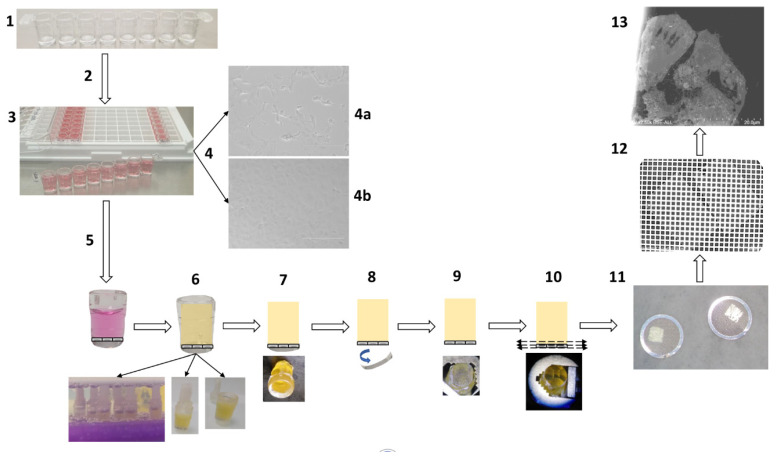
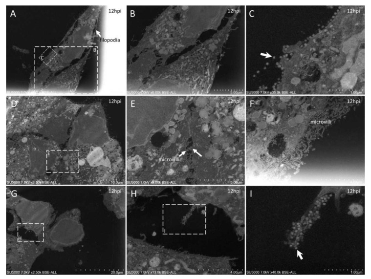
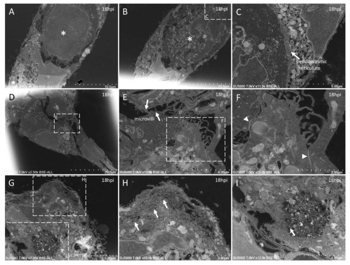
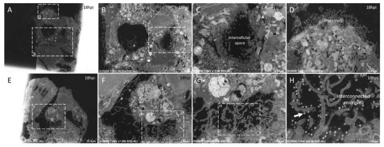
Despite the development of new diagnostic methods, co-culture, based on sample inoculation of cell monolayers coupled with electron microscopy (EM) observation, remains the gold standard in virology. Indeed, co-culture allows for the study of cell morphology (infected and not infected), the ultrastructure of the inoculated virus, and the different steps of the virus infectious cycle. Most EM methods for studying virus cycles are applied after infected cells are produced in large quantities and detached to obtain a pellet. Here, cell culture was performed in sterilized, collagen-coated single-break strip wells. After one day in culture, cells were infected with SARS-CoV-2. Wells of interest were fixed at different time points, from 2 to 36 h post-infection. Microwave-assisted resin embedding was accomplished directly in the wells in 4 h. Finally, ultra-thin sections were cut directly through the infected-cell monolayers. Our methodology requires, in total, less than four days for preparing and observing cells. Furthermore, by observing undetached infected cell monolayers, we were able to observe new ultrastructural findings, such as cell-cell interactions and baso-apical cellular organization related to the virus infectious cycle. Our innovative methodology thus not only saves time for preparation but also adds precision and new knowledge about viral infection, as shown here for SARS-CoV-2.

摘要

尽管新的诊断方法不断发展,但基于细胞单层接种并结合电子显微镜(EM)观察的共培养,仍然是病毒学的金标准。事实上,共培养能够研究细胞形态(感染和未感染的)、接种病毒的超微结构以及病毒感染周期的不同步骤。大多数用于研究病毒周期的EM方法是在大量生产并分离感染细胞以获得沉淀后应用的。在此,细胞培养在经过消毒、涂有胶原蛋白的单孔条孔板中进行。培养一天后,用严重急性呼吸综合征冠状病毒2(SARS-CoV-2)感染细胞。在感染后2至36小时的不同时间点固定感兴趣的孔板。4小时内在孔板中直接完成微波辅助树脂包埋。最后,直接穿过感染细胞单层切出超薄切片。我们的方法总共只需不到四天的时间来制备和观察细胞。此外,通过观察未分离的感染细胞单层,我们能够观察到新的超微结构发现,例如与病毒感染周期相关的细胞间相互作用和基底-顶端细胞组织。因此,我们的创新方法不仅节省了制备时间,还增加了关于病毒感染的精确性和新知识,如本文针对SARS-CoV-2所展示的。

https://cdn.ncbi.nlm.nih.gov/pmc/blobs/3013/8228702/6ba91cc25e7d/microorganisms-09-01194-g007.jpg
https://cdn.ncbi.nlm.nih.gov/pmc/blobs/3013/8228702/95b6898377c5/microorganisms-09-01194-g001.jpg
https://cdn.ncbi.nlm.nih.gov/pmc/blobs/3013/8228702/208cd9f11366/microorganisms-09-01194-g002.jpg
https://cdn.ncbi.nlm.nih.gov/pmc/blobs/3013/8228702/b83b2cc61fed/microorganisms-09-01194-g003.jpg
https://cdn.ncbi.nlm.nih.gov/pmc/blobs/3013/8228702/2cb9d0b6e47a/microorganisms-09-01194-g004.jpg
https://cdn.ncbi.nlm.nih.gov/pmc/blobs/3013/8228702/39891f7b0783/microorganisms-09-01194-g005.jpg
https://cdn.ncbi.nlm.nih.gov/pmc/blobs/3013/8228702/25b08134d46b/microorganisms-09-01194-g006.jpg
https://cdn.ncbi.nlm.nih.gov/pmc/blobs/3013/8228702/6ba91cc25e7d/microorganisms-09-01194-g007.jpg

相似文献

1
Innovative Approach to Fast Electron Microscopy Using the Example of a Culture of Virus-Infected Cells: An Application to SARS-CoV-2.以病毒感染细胞培养物为例的快速电子显微镜创新方法:在严重急性呼吸综合征冠状病毒2(SARS-CoV-2)中的应用
Microorganisms. 2021 May 31;9(6):1194. doi: 10.3390/microorganisms9061194.
2
Microwave-assisted tissue processing for same-day EM-diagnosis of potential bioterrorism and clinical samples.用于潜在生物恐怖主义和临床样本当日电镜诊断的微波辅助组织处理
Micron. 2006;37(6):577-90. doi: 10.1016/j.micron.2005.11.015.
3
Concentration of SARS-CoV-2-Infected Cell Culture Supernatants for Detection of Virus-like Particles by Scanning Electron Microscopy.通过扫描电子显微镜检测病毒样颗粒的 SARS-CoV-2 感染细胞培养上清液的浓缩。
Viruses. 2022 Oct 28;14(11):2388. doi: 10.3390/v14112388.
4
The Strengths of Scanning Electron Microscopy in Deciphering SARS-CoV-2 Infectious Cycle.扫描电子显微镜在解析新型冠状病毒感染周期方面的优势
Front Microbiol. 2020 Aug 19;11:2014. doi: 10.3389/fmicb.2020.02014. eCollection 2020.
5
SARS-CoV-2 infection diagnosed only by cell culture isolation before the local outbreak in an Italian seven-week-old suckling baby.在当地暴发前,通过细胞培养分离仅诊断出一例意大利 7 周龄哺乳婴儿感染 SARS-CoV-2。
Int J Infect Dis. 2020 Jul;96:387-389. doi: 10.1016/j.ijid.2020.05.035. Epub 2020 May 14.
6
Preparation of herpes simplex virus-infected primary neurons for transmission electron microscopy.用于透射电子显微镜观察的单纯疱疹病毒感染原代神经元的制备
Methods Mol Biol. 2014;1144:223-34. doi: 10.1007/978-1-4939-0428-0_15.
7
Preparation of Herpes Simplex Virus-Infected Primary Neurons for Transmission Electron Microscopy.用于透射电子显微镜观察的单纯疱疹病毒感染原代神经元的制备
Methods Mol Biol. 2020;2060:343-354. doi: 10.1007/978-1-4939-9814-2_20.
8
Two Detailed Plaque Assay Protocols for the Quantification of Infectious SARS-CoV-2.两种用于定量检测传染性严重急性呼吸综合征冠状病毒2(SARS-CoV-2)的详细噬斑测定方案。
Curr Protoc Microbiol. 2020 Jun;57(1):ecpmc105. doi: 10.1002/cpmc.105.
9
Application of transmission electron microscopy to the clinical study of viral and bacterial infections: present and future.透射电子显微镜在病毒和细菌感染临床研究中的应用:现状与未来
Micron. 2006;37(2):91-106. doi: 10.1016/j.micron.2005.10.001. Epub 2005 Nov 2.
10
3D Electron Microscopy (EM) and Correlative Light Electron Microscopy (CLEM) Methods to Study Virus-Host Interactions.用于研究病毒-宿主相互作用的3D电子显微镜(EM)和相关光电子显微镜(CLEM)方法。
Methods Mol Biol. 2018;1836:213-236. doi: 10.1007/978-1-4939-8678-1_11.

引用本文的文献

1
Monkeypox Virus Subverts the Inflammatory Response of Macrophages at the Maternal-Fetal Interface.猴痘病毒破坏母婴界面巨噬细胞的炎症反应。
J Med Virol. 2025 May;97(5):e70412. doi: 10.1002/jmv.70412.
2
escapes LAPosome and modulates macrophage response in a xenophagy-dependent manner.逃脱脂质体自噬体(LAPosome)并以自噬依赖性方式调节巨噬细胞反应。
Autophagy Rep. 2025 Mar 11;4(1):2475527. doi: 10.1080/27694127.2025.2475527. eCollection 2025.
3
Microscopic observations of SARS-CoV-2 like particles in different oral samples.不同口腔样本中类似 SARS-CoV-2 的微粒的微观观察。

本文引用的文献

1
Difficulties in Differentiating Coronaviruses from Subcellular Structures in Human Tissues by Electron Microscopy.电子显微镜下鉴别人组织中冠状病毒与亚细胞结构的困难。
Emerg Infect Dis. 2021 Apr;27(4):1023-1031. doi: 10.3201/eid2704.204337. Epub 2021 Feb 18.
2
Morphometry of SARS-CoV and SARS-CoV-2 particles in ultrathin plastic sections of infected Vero cell cultures.感染的 Vero 细胞培养物超薄塑料切片中 SARS-CoV 和 SARS-CoV-2 颗粒的形态测量。
Sci Rep. 2021 Feb 10;11(1):3515. doi: 10.1038/s41598-021-82852-7.
3
Ultrastructural modifications induced by SARS-CoV-2 in Vero cells: a kinetic analysis of viral factory formation, viral particle morphogenesis and virion release.
Eur J Oral Sci. 2022 Dec;130(6):e12903. doi: 10.1111/eos.12903. Epub 2022 Nov 20.
4
Concentration of SARS-CoV-2-Infected Cell Culture Supernatants for Detection of Virus-like Particles by Scanning Electron Microscopy.通过扫描电子显微镜检测病毒样颗粒的 SARS-CoV-2 感染细胞培养上清液的浓缩。
Viruses. 2022 Oct 28;14(11):2388. doi: 10.3390/v14112388.
5
Limited permissibility of ENL-R and Mv-1-Lu mink cell lines to SARS-CoV-2.ENL-R和Mv-1-Lu貂细胞系对严重急性呼吸综合征冠状病毒2(SARS-CoV-2)的允许性有限。
Front Microbiol. 2022 Oct 12;13:1003824. doi: 10.3389/fmicb.2022.1003824. eCollection 2022.
6
Control of /E-Cadherin Gene Expression and Release of a Soluble Form of E-Cadherin in SARS-CoV-2 Infected Caco-2 Intestinal Cells: Physiopathological Consequences for the Intestinal Forms of COVID-19.SARS-CoV-2 感染的 Caco-2 肠细胞中/E-钙黏蛋白基因表达的控制和可溶性 E-钙黏蛋白的释放:COVID-19 肠道形式的病理生理后果。
Front Cell Infect Microbiol. 2022 May 4;12:798767. doi: 10.3389/fcimb.2022.798767. eCollection 2022.
SARS-CoV-2 对 Vero 细胞的超微结构改变:病毒工厂形成、病毒粒子形态发生和病毒粒子释放的动力学分析。
Cell Mol Life Sci. 2021 Apr;78(7):3565-3576. doi: 10.1007/s00018-020-03745-y. Epub 2021 Jan 15.
4
The Strengths of Scanning Electron Microscopy in Deciphering SARS-CoV-2 Infectious Cycle.扫描电子显微镜在解析新型冠状病毒感染周期方面的优势
Front Microbiol. 2020 Aug 19;11:2014. doi: 10.3389/fmicb.2020.02014. eCollection 2020.
5
Morphogenesis and cytopathic effect of SARS-CoV-2 infection in human airway epithelial cells.SARS-CoV-2 感染人呼吸道上皮细胞的形态发生和细胞病变效应。
Nat Commun. 2020 Aug 6;11(1):3910. doi: 10.1038/s41467-020-17796-z.
6
A molecular pore spans the double membrane of the coronavirus replication organelle.一种分子孔跨越冠状病毒复制细胞器的双层膜。
Science. 2020 Sep 11;369(6509):1395-1398. doi: 10.1126/science.abd3629. Epub 2020 Aug 6.
7
SARS-coronavirus-2 replication in Vero E6 cells: replication kinetics, rapid adaptation and cytopathology.严重急性呼吸综合征冠状病毒-2 在 Vero E6 细胞中的复制:复制动力学、快速适应和细胞病变。
J Gen Virol. 2020 Sep;101(9):925-940. doi: 10.1099/jgv.0.001453.
8
Viral RNA load as determined by cell culture as a management tool for discharge of SARS-CoV-2 patients from infectious disease wards.通过细胞培养测定病毒 RNA 载量作为管理工具,以将 SARS-CoV-2 患者从传染病病房中出院。
Eur J Clin Microbiol Infect Dis. 2020 Jun;39(6):1059-1061. doi: 10.1007/s10096-020-03913-9. Epub 2020 Apr 27.
9
A Novel Coronavirus from Patients with Pneumonia in China, 2019.2019 年中国肺炎患者中的一种新型冠状病毒。
N Engl J Med. 2020 Feb 20;382(8):727-733. doi: 10.1056/NEJMoa2001017. Epub 2020 Jan 24.
10
The viral replication organelles within cells studied by electron microscopy.电子显微镜下研究细胞内的病毒复制细胞器。
Adv Virus Res. 2019;105:1-33. doi: 10.1016/bs.aivir.2019.07.005. Epub 2019 Aug 19.