Osman Mazen, Akkus Zeynettin, Jevremovic Dragan, Nguyen Phuong L, Roh Dana, Al-Kali Aref, Patnaik Mrinal M, Nanaa Ahmad, Rizk Samia, Salama Mohamed E
Division of Anatomic and Clinical Pathology, Mayo Clinic, Rochester, MN 55905, USA.
Division of Cardiovascular Diseases, Mayo Clinic, Rochester, MN 55905, USA.
J Clin Med. 2021 May 24;10(11):2264. doi: 10.3390/jcm10112264.
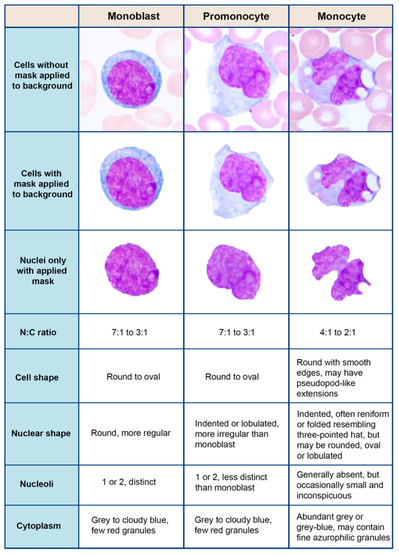
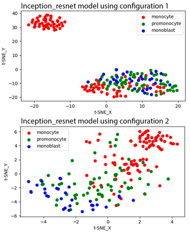
The accurate diagnosis of chronic myelomonocytic leukemia (CMML) and acute myeloid leukemia (AML) subtypes with monocytic differentiation relies on the proper identification and quantitation of blast cells and blast-equivalent cells, including promonocytes. This distinction can be quite challenging given the cytomorphologic and immunophenotypic similarities among the monocytic cell precursors. The aim of this study was to assess the performance of convolutional neural networks (CNN) in separating monocytes from their precursors (i.e., promonocytes and monoblasts). We collected digital images of 935 monocytic cells that were blindly reviewed by five experienced morphologists and assigned into three subtypes: monocyte, promonocyte, and blast. The consensus between reviewers was considered as a ground truth reference label for each cell. In order to assess the performance of CNN models, we divided our data into training (70%), validation (10%), and test (20%) datasets, as well as applied fivefold cross validation. The CNN models did not perform well for predicting three monocytic subtypes, but their performance was significantly improved for two subtypes (monocyte vs. promonocytes + blasts). Our findings (1) support the concept that morphologic distinction between monocytic cells of various differentiation level is difficult; (2) suggest that combining blasts and promonocytes into a single category is desirable for improved accuracy; and (3) show that CNN models can reach accuracy comparable to human reviewers (0.78 ± 0.10 vs. 0.86 ± 0.05). As far as we know, this is the first study to separate monocytes from their precursors using CNN.
慢性粒单核细胞白血病(CMML)和具有单核细胞分化的急性髓系白血病(AML)亚型的准确诊断依赖于对原始细胞和原始细胞等效细胞(包括原单核细胞)的正确识别和定量。鉴于单核细胞前体之间的细胞形态学和免疫表型相似性,这种区分颇具挑战性。本研究的目的是评估卷积神经网络(CNN)在区分单核细胞与其前体(即原单核细胞和单核母细胞)方面的性能。我们收集了935个单核细胞的数字图像,由五位经验丰富的形态学家进行盲法评估,并分为三个亚型:单核细胞、原单核细胞和原始细胞。评审者之间的共识被视为每个细胞的真实参考标签。为了评估CNN模型的性能,我们将数据分为训练集(70%)、验证集(10%)和测试集(20%),并应用五折交叉验证。CNN模型在预测三种单核细胞亚型方面表现不佳,但其在两种亚型(单核细胞与原单核细胞+原始细胞)上的性能显著提高。我们的研究结果(1)支持不同分化水平的单核细胞之间形态学区分困难的概念;(2)表明将原始细胞和原单核细胞合并为一个类别以提高准确性是可取的;(3)表明CNN模型可以达到与人类评审者相当的准确率(0.78±0.10对0.86±0.05)。据我们所知,这是第一项使用CNN区分单核细胞及其前体的研究。