Medical Isotopes Research Center and Department of Radiation Medicine, School of Basic Medical Sciences, Peking University Health Science Center, Beijing, 100191, China.
Key Laboratory of Carcinogenesis and Translational Research (Ministry of Education/Beijing), Department of Nuclear Medicine, Peking University Cancer Hospital & Institute, Beijing, 100142, China.
J Nanobiotechnology. 2021 Jun 10;19(1):175. doi: 10.1186/s12951-021-00924-2.
Adoptive T cell transfer-based immunotherapy yields unsatisfactory results in the treatment of solid tumors, partially owing to limited tumor infiltration and the immunosuppressive microenvironment in solid tumors. Therefore, strategies for the noninvasive tracking of adoptive T cells are critical for monitoring tumor infiltration and for guiding the development of novel combination therapies.
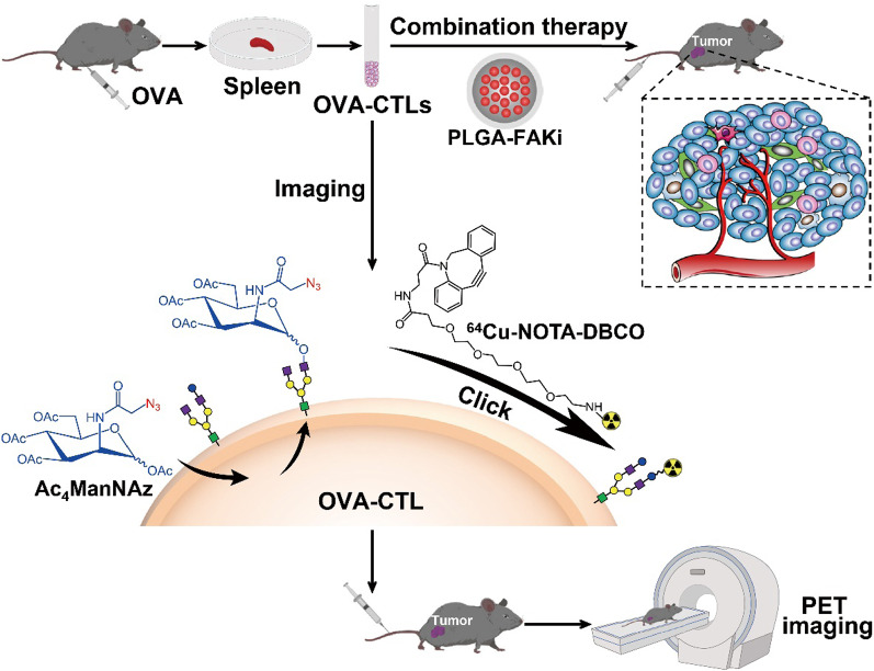
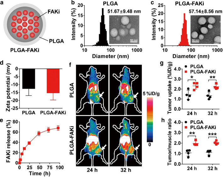
We developed a radiolabeling method for cytotoxic T lymphocytes (CTLs) that comprises metabolically labeling the cell surface glycans with azidosugars and then covalently conjugating them with Cu-1,4,7-triazacyclononanetriacetic acid-dibenzo-cyclooctyne (Cu-NOTA-DBCO) using bioorthogonal chemistry. Cu-labeled control-CTLs and ovalbumin-specific CTLs (OVA-CTLs) were tracked using positron emission tomography (PET) in B16-OVA tumor-bearing mice. We also investigated the effects of focal adhesion kinase (FAK) inhibition on the antitumor efficacy of OVA-CTLs using a poly(lactic-co-glycolic) acid (PLGA)-encapsulated nanodrug (PLGA-FAKi).
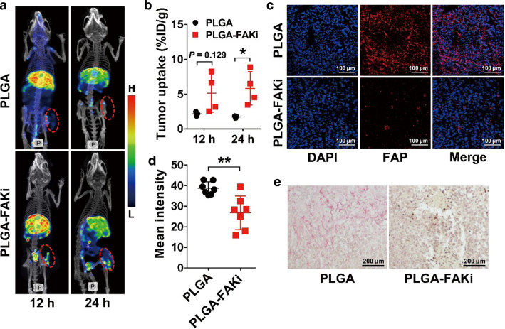
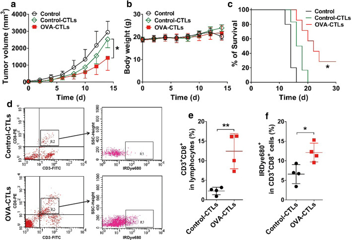
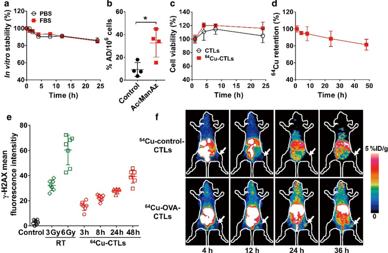
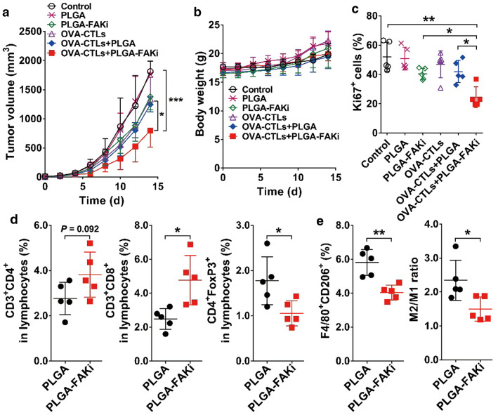
CTLs can be stably radiolabeled with Cu with a minimal effect on cell viability. PET imaging of Cu-OVA-CTLs enables noninvasive mapping of their in vivo behavior. Moreover, Cu-OVA-CTLs PET imaging revealed that PLGA-FAKi induced a significant increase in OVA-CTL infiltration into tumors, suggesting the potential for a combined therapy comprising OVA-CTLs and PLGA-FAKi. Further combination therapy studies confirmed that the PLGA-FAKi nanodrug markedly improved the antitumor effects of adoptive OVA-CTLs transfer by multiple mechanisms.
These findings demonstrated that metabolic radiolabeling followed by PET imaging can be used to sensitively profile the early-stage migration and tumor-targeting efficiency of adoptive T cells in vivo. This strategy presents opportunities for predicting the efficacy of cell-based adoptive therapies and for guiding combination regimens.
过继性 T 细胞转移的免疫疗法在治疗实体瘤方面的效果并不理想,部分原因是实体瘤中 T 细胞浸润有限和免疫抑制微环境。因此,非侵入性跟踪过继性 T 细胞的策略对于监测肿瘤浸润和指导新型联合治疗的发展至关重要。
我们开发了一种用于细胞毒性 T 淋巴细胞(CTLs)的放射性标记方法,该方法包括通过代谢将糖基化的细胞表面糖用叠氮化物标记,然后使用生物正交化学将其与 Cu-1,4,7-三氮杂环壬烷三乙酸-二苯并环辛炔(Cu-NOTA-DBCO)共价连接。使用正电子发射断层扫描(PET)在 B16-OVA 肿瘤荷瘤小鼠中追踪 Cu 标记的对照 CTL 和卵清蛋白特异性 CTL(OVA-CTL)。我们还研究了使用聚乳酸-羟基乙酸共聚物(PLGA)包封的纳米药物(PLGA-FAKi)抑制粘着斑激酶(FAK)对 OVA-CTL 抗肿瘤功效的影响。
CTL 可以稳定地用 Cu 放射性标记,对细胞活力的影响最小。Cu-OVA-CTL 的 PET 成像能够对其体内行为进行非侵入性的映射。此外,Cu-OVA-CTL 的 PET 成像显示,PLGA-FAKi 可显著增加 OVA-CTL 浸润肿瘤,表明 OVA-CTL 和 PLGA-FAKi 联合治疗具有潜力。进一步的联合治疗研究证实,PLGA-FAKi 纳米药物通过多种机制显著提高了过继性 OVA-CTL 转移的抗肿瘤效果。
这些发现表明,代谢放射性标记后进行 PET 成像可以用于敏感地分析体内过继性 T 细胞的早期迁移和肿瘤靶向效率。这种策略为预测细胞基础过继性治疗的疗效和指导联合治疗方案提供了机会。