Department of Veterinary Medicine, Università degli Studi di Milano, Lodi, Italy.
Centro Clinico-Veterinario e Zootecnico-Sperimentale, Università degli Studi di Milano, Lodi, Italy.
Vet Radiol Ultrasound. 2021 Sep;62(5):602-609. doi: 10.1111/vru.12995. Epub 2021 Jun 15.
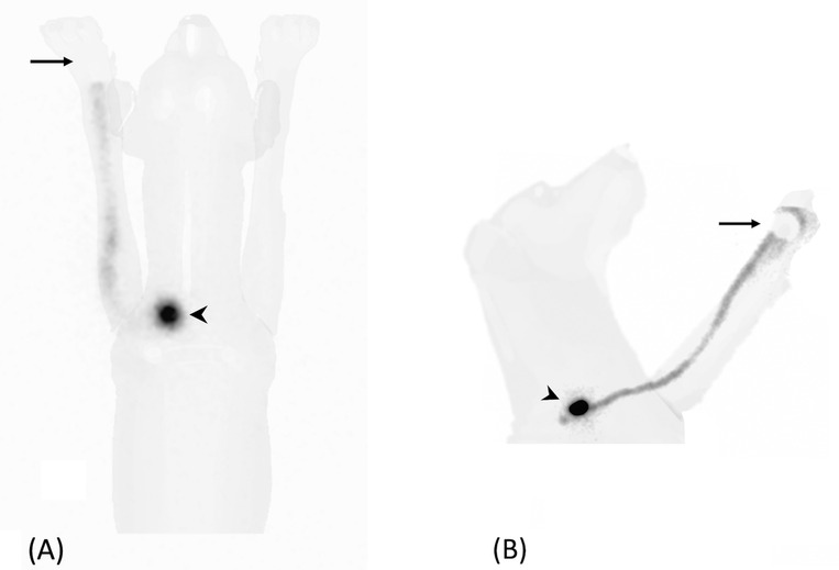
Sentinel lymph node (SLN) mapping is the current gold standard for the oncological staging of solid malignancies in humans. This prospective observational study describes the feasibility and the limits of preoperative lymphoscintigraphy for SLN detection in dogs with spontaneous malignancies and the improvements in staging accuracy. Client-owned dogs with confirmed malignant neoplasia and absence of distant metastasis were prospectively enrolled. Lymphoscintigraphy was performed after the peritumoral injection of Technetium-99m labeled nanocolloids. Regional dynamic and static images were acquired, with and without masking of the injection site with a lead shield. The dogs were then subjected to surgery for tumor excision and SLN extirpation. Intraoperative SLN detection was performed by combining methylene blue dye and a dedicated gamma probe. Overall, 51 dogs with a total of 60 solid malignant tumors were enrolled. Lymphoscintigraphy identified at least one SLN in 57 of 60 cases (95%). The SLN did not always correspond to the regional lymph node (35/57, 61.4%). The use of a lead shield, masking the injection site, markedly improved the SLN visibility. The median time of SLN appearance was 11.4 ± 9.3 min. No side effects were observed. Preoperative lymphoscintigraphy allows for SLN detection in dogs and can improve staging accuracy by either identifying the SLN in a different lymphosome than clinically expected or discriminating the draining node in uncertain cases. The combined use of preoperative and intraoperative techniques is recommended to increase the SLN detection rate.
前哨淋巴结 (SLN) 作图是目前人类实体恶性肿瘤肿瘤分期的金标准。这项前瞻性观察研究描述了术前淋巴闪烁显像术检测自发性恶性肿瘤犬 SLN 的可行性和局限性,以及分期准确性的提高。经确认患有恶性肿瘤且无远处转移的患犬被前瞻性纳入。在肿瘤周围注射锝-99m 标记的纳米胶体后,进行淋巴闪烁显像术。进行区域动态和静态图像采集,同时用铅屏蔽屏蔽注射部位。然后,对犬进行肿瘤切除术和 SLN 切除术。术中通过结合亚甲蓝染料和专用伽马探针进行 SLN 检测。共有 51 只犬患有 60 个实体恶性肿瘤,总体上纳入研究。60 例中,57 例(95%)至少检出 1 个 SLN。SLN 并不总是对应于区域性淋巴结(35/57,61.4%)。使用铅屏蔽,屏蔽注射部位,可显著提高 SLN 的可视性。SLN 出现的中位时间为 11.4±9.3 分钟。未观察到副作用。术前淋巴闪烁显像术可用于犬的 SLN 检测,并通过在临床预期以外的不同淋巴结中识别 SLN 或在不确定的情况下区分引流淋巴结,从而提高分期准确性。建议联合使用术前和术中技术以提高 SLN 检测率。