• 文献检索
  • 文档翻译
  • 深度研究
  • 学术资讯
  • Suppr Zotero 插件Zotero 插件
  • 邀请有礼
  • 套餐&价格
  • 历史记录
应用&插件
Suppr Zotero 插件Zotero 插件浏览器插件Mac 客户端Windows 客户端微信小程序
定价
高级版会员购买积分包购买API积分包
服务
文献检索文档翻译深度研究API 文档MCP 服务
关于我们
关于 Suppr公司介绍联系我们用户协议隐私条款
关注我们

Suppr 超能文献

核心技术专利:CN118964589B侵权必究
粤ICP备2023148730 号-1Suppr @ 2026

文献检索

告别复杂PubMed语法,用中文像聊天一样搜索,搜遍4000万医学文献。AI智能推荐,让科研检索更轻松。

立即免费搜索

文件翻译

保留排版,准确专业,支持PDF/Word/PPT等文件格式,支持 12+语言互译。

免费翻译文档

深度研究

AI帮你快速写综述,25分钟生成高质量综述,智能提取关键信息,辅助科研写作。

立即免费体验

轻度 COVID-19 感染患者的中央凹和脉络膜厚度如何受到影响?

How are central foveal and choroidal thickness affected in patients with mild COVID-19 infection?

机构信息

Department of Ophthalmology, Elazığ City Hospital, Elazığ, Turkey.

出版信息

Bosn J Basic Med Sci. 2021 Dec 1;21(6):782-786. doi: 10.17305/bjbms.2021.5840.

DOI:10.17305/bjbms.2021.5840
PMID:34157253
原文链接:https://pmc.ncbi.nlm.nih.gov/articles/PMC8554697/
Abstract

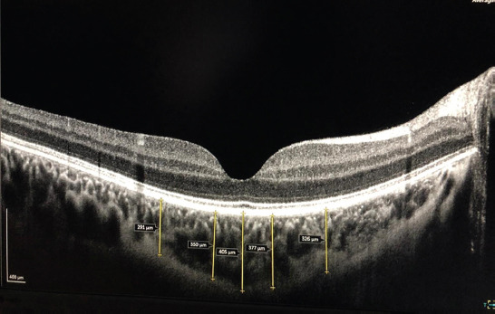
The aim of this study was to evaluate the effects of  COVID-19 on central foveal and choroidal thicknesses. Thirty-two patients with a positive SARS-CoV-2 PCR test who received outpatient treatment within the previous two months and 32 healthy controls were included in the study. Patients requiring hospitalization due to COVID-19 as well as the patients who received either intensive care support and/or antiplatelet therapy, smokers, or patients with systemic or ocular diseases were excluded from the study. After full ophthalmological examination, central foveal and choroidal thicknesses were evaluated by using optical coherence tomography. Statistical analysis of the study data demonstrated no significant difference between the groups in terms of age or gender (p>0.05). There was also no statistically significant difference between the groups in terms of central foveal thickness, central choroidal thickness, or nasal 500, nasal 1500, temporal 500, or temporal 500-micron distances (p>0.05 for all parameters). Choroidal and retinal thicknesses were not affected in patients with recent mild COVID 19 without comorbidities.

摘要

本研究旨在评估 COVID-19 对中央凹和脉络膜厚度的影响。研究纳入了 32 名在过去两个月内接受门诊治疗且 SARS-CoV-2 PCR 检测呈阳性的患者和 32 名健康对照者。因 COVID-19 需要住院治疗以及接受重症监护支持和/或抗血小板治疗、吸烟者或患有系统性或眼部疾病的患者被排除在研究之外。在进行全面的眼科检查后,使用光学相干断层扫描评估中央凹和脉络膜厚度。对研究数据的统计学分析显示,两组在年龄或性别方面无显著差异(p>0.05)。两组在中央凹厚度、脉络膜中央厚度或鼻侧 500、鼻侧 1500、颞侧 500 或颞侧 500 微米距离方面也无统计学差异(所有参数 p>0.05)。无合并症的近期轻度 COVID-19 患者的脉络膜和视网膜厚度不受影响。

https://cdn.ncbi.nlm.nih.gov/pmc/blobs/fa41/8554697/4e2a4bcce157/BJBMS-21-782-g001.jpg

相似文献

1
How are central foveal and choroidal thickness affected in patients with mild COVID-19 infection?轻度 COVID-19 感染患者的中央凹和脉络膜厚度如何受到影响?
Bosn J Basic Med Sci. 2021 Dec 1;21(6):782-786. doi: 10.17305/bjbms.2021.5840.
2
Evaluation of choroidal thickness with OCT in COVID-19 patients with high D-dimer levels.评估 COVID-19 患者高 D-二聚体水平的脉络膜厚度与 OCT 的关系。
Sci Rep. 2022 Oct 18;12(1):16826. doi: 10.1038/s41598-022-21579-5.
3
Choroidal and central foveal thickness in patients with scleroderma and its systemic associations.硬皮病患者的脉络膜及中央凹厚度及其全身关联
Clin Exp Optom. 2017 Nov;100(6):656-662. doi: 10.1111/cxo.12498. Epub 2016 Nov 18.
4
Evaluation of the Choroid, Fovea, and Retinal Nerve Fiber Layer in Patients with Rheumatoid Arthritis.类风湿关节炎患者脉络膜、黄斑和视网膜神经纤维层的评估
Ocul Immunol Inflamm. 2017 Apr;25(2):210-214. doi: 10.3109/09273948.2015.1095303. Epub 2015 Dec 30.
5
Analysis of choroidal and central foveal thicknesses in acute anterior uveitis by enhanced-depth imaging optical coherence tomography.增强深度成像光学相干断层扫描分析急性前葡萄膜炎患者的脉络膜和中心凹厚度
BMC Ophthalmol. 2017 Dec 1;17(1):225. doi: 10.1186/s12886-017-0628-7.
6
Analysis of spectral-domain optical coherence tomography in preterm children: retinal layer thickness and choroidal thickness profiles.早产儿的谱域光相干断层扫描分析:视网膜层厚度和脉络膜厚度曲线。
Invest Ophthalmol Vis Sci. 2012 Oct 17;53(11):7201-7. doi: 10.1167/iovs.12-10599.
7
Effect of smoking on choroidal thickness in healthy smokers.吸烟对健康吸烟者脉络膜厚度的影响。
Curr Eye Res. 2014 May;39(5):504-11. doi: 10.3109/02713683.2013.850099. Epub 2013 Nov 11.
8
QUANTITATIVE ANALYSIS OF STRUCTURAL ALTERATIONS IN THE CHOROID OF PATIENTS WITH ACTIVE BEHÇET UVEITIS.活动性 Behcet 葡萄膜炎患者脉络膜结构改变的定量分析。
Retina. 2018 Apr;38(4):828-840. doi: 10.1097/IAE.0000000000001587.
9
Foveal and parafoveal choroidal thickness pattern measuring by swept source optical coherence tomography.应用扫频源光学相干断层扫描测量中心凹及旁中心凹脉络膜厚度。
Eye (Lond). 2019 Sep;33(9):1443-1451. doi: 10.1038/s41433-019-0404-4. Epub 2019 Apr 8.
10
Choroidal thickness in psoriasis.银屑病中的脉络膜厚度。
Int Ophthalmol. 2017 Feb;37(1):173-177. doi: 10.1007/s10792-016-0252-1. Epub 2016 May 11.

引用本文的文献

1
Effect of the COVID-19 pandemic on Vogt-Koyanagi-Harada disease.COVID-19 大流行对 Vogt-Koyanagi-Harada 病的影响。
Sci Rep. 2024 Jun 8;14(1):13211. doi: 10.1038/s41598-024-63957-1.
2
New findings on retinal microvascular changes in patients with primary COVID-19 infection: a longitudinal study.原发性 COVID-19 感染患者视网膜微血管变化的新发现:一项纵向研究。
Front Immunol. 2024 May 21;15:1404785. doi: 10.3389/fimmu.2024.1404785. eCollection 2024.
3
Ocular manifestations of COVID-19: A systematic review of current evidence.

本文引用的文献

1
Retinal microvascular abnormalities in patients after COVID-19 depending on disease severity.新冠病毒感染后患者的视网膜血管异常与疾病严重程度有关。
Br J Ophthalmol. 2022 Apr;106(4):559-563. doi: 10.1136/bjophthalmol-2020-317953. Epub 2020 Dec 16.
2
Retinal findings in hospitalised patients with severe COVID-19.住院严重 COVID-19 患者的视网膜表现。
Br J Ophthalmol. 2022 Jan;106(1):102-105. doi: 10.1136/bjophthalmol-2020-317576. Epub 2020 Oct 16.
3
Retinal findings in patients with COVID-19: Results from the SERPICO-19 study.
新型冠状病毒肺炎的眼部表现:当前证据的系统评价
Prev Med Rep. 2024 Jan 14;38:102608. doi: 10.1016/j.pmedr.2024.102608. eCollection 2024 Feb.
4
Retinal tissue and microvasculature loss in COVID-19 infection.新冠病毒感染导致的视网膜组织和微血管损伤。
Sci Rep. 2023 Mar 29;13(1):5100. doi: 10.1038/s41598-023-31835-x.
5
Evaluation of choroidal thickness with OCT in COVID-19 patients with high D-dimer levels.评估 COVID-19 患者高 D-二聚体水平的脉络膜厚度与 OCT 的关系。
Sci Rep. 2022 Oct 18;12(1):16826. doi: 10.1038/s41598-022-21579-5.
6
Retrobulbar ocular blood flow and choroidal vascular changes in patients recovering from COVID-19 infection.新冠病毒感染后恢复期患者的眼后段血流和脉络膜血管变化。
Photodiagnosis Photodyn Ther. 2022 Sep;39:102976. doi: 10.1016/j.pdpdt.2022.102976. Epub 2022 Jun 17.
7
Spectral-domain optical coherence tomography assessment of retinal and choroidal changes in patients with coronavirus disease 2019: a case-control study.2019冠状病毒病患者视网膜和脉络膜变化的光谱域光学相干断层扫描评估:一项病例对照研究
J Ophthalmic Inflamm Infect. 2022 Jun 18;12(1):18. doi: 10.1186/s12348-022-00297-z.
新型冠状病毒肺炎患者的视网膜表现:SERPICO-19研究结果
EClinicalMedicine. 2020 Oct;27:100550. doi: 10.1016/j.eclinm.2020.100550. Epub 2020 Sep 20.
4
SARS-CoV-2 Targeting the Retina: Host-virus Interaction and Possible Mechanisms of Viral Tropism.SARS-CoV-2 靶向视网膜:宿主-病毒相互作用及病毒趋向性的可能机制。
Ocul Immunol Inflamm. 2020 Nov 16;28(8):1301-1304. doi: 10.1080/09273948.2020.1799037. Epub 2020 Sep 18.
5
Papillophlebitis in a COVID-19 patient: Inflammation and hypercoagulable state.COVID-19 患者的乳头静脉炎:炎症和高凝状态。
Eur J Ophthalmol. 2022 Jan;32(1):NP168-NP172. doi: 10.1177/1120672120947591. Epub 2020 Jul 30.
6
COVID-19 retinal microangiopathy as an in vivo biomarker of systemic vascular disease?COVID-19 视网膜微血管病变是否可作为系统性血管疾病的体内生物标志物?
J Intern Med. 2021 Jan;289(1):116-120. doi: 10.1111/joim.13156. Epub 2020 Jul 30.
7
Concerns about the interpretation of OCT and fundus findings in COVID-19 patients in recent Lancet publication.近期《柳叶刀》发表的文章对新冠病毒肺炎患者光学相干断层扫描(OCT)及眼底检查结果的解读提出了担忧。
Eye (Lond). 2020 Dec;34(12):2153-2154. doi: 10.1038/s41433-020-1084-9. Epub 2020 Jul 9.
8
Paracentral acute middle maculopathy and acute macular neuroretinopathy following SARS-CoV-2 infection.新型冠状病毒感染后的中心旁急性黄斑病变和急性黄斑神经视网膜病变
Eye (Lond). 2020 Dec;34(12):2352-2353. doi: 10.1038/s41433-020-1069-8. Epub 2020 Jul 3.
9
Bilateral anterior uveitis as a part of a multisystem inflammatory syndrome secondary to COVID-19 infection.双侧前葡萄膜炎作为新型冠状病毒肺炎(COVID-19)感染继发的多系统炎症综合征的一部分。
J Med Virol. 2021 Jan;93(1):139-140. doi: 10.1002/jmv.26229. Epub 2020 Sep 30.
10
Unique case of central retinal artery occlusion secondary to COVID-19 disease.新型冠状病毒肺炎继发中央视网膜动脉阻塞的罕见病例。
IDCases. 2020;21:e00867. doi: 10.1016/j.idcr.2020.e00867. Epub 2020 Jun 18.