Campos Raquel M P, Barbosa-Silva Maria Carolina, Ribeiro-Resende Victor T
Instituto de Biofísica Carlos Chagas Filho, Laboratório de Neuroquímica, Universidade Federal do Rio de Janeiro, Rio de Janeiro, RJ 21941-902, Brazil.
Núcleo Multidisciplinar de Pesquisa em Biologia (Numpex-Bio), Campus de Duque de Caxias Geraldo Guerra Cidade, Universidade Federal do Rio de Janeiro, Duque de Caxias, RJ 25255-030, Brazil.
IBRO Neurosci Rep. 2021 May 16;10:225-235. doi: 10.1016/j.ibneur.2021.05.002. eCollection 2021 Jun.
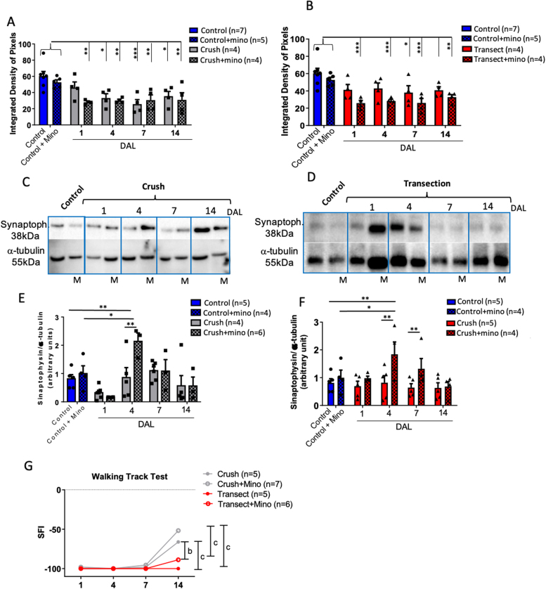
In an injury to the peripheral nervous system, the spinal cord and brain structure reorganize connections to optimize the function of the remaining parts. Many cell events are triggered in the spinal cord to support changes in the synaptic connections around motoneurons, where old connections are removed, and new ones created. Microglial cells are primitive macrophages that invade the central nervous system in early stages of neurodevelopment and have several functions, such as eliminating synapses. We investigated the synaptic plasticity after different types of peripheral (sciatic) nerve injury (crush or total transection), as well as the behavior of microglial cells for 2 weeks after a peripheral lesion. As expected, sciatic-nerve injury reduced motor performance in mice, but crushed animals regained partial motor control. Because of sciatic-nerve injury, pre-synaptic inputs decreased around the motoneurons in the ventro-lateral horn, while microglial cells increased around these cells. Microglial cells also exhibited altered morphology in both types of peripheral lesion, indicating a similar underlying mechanism of plasticity. To investigate the involvement of microglia in this scenario, microglial activation was modulated by daily administration of minocycline. The minocycline treatment directly affected the microglial response and impacted the synapse rearrangement in the spinal cord. Together, these results demonstrate that microglia cells are involved in synaptic plasticity in the lumbar spinal cord in both nerve-injury scenarios.
Here, we demonstrated that acute plasticity in the lumbar spinal cord (LSC) did not differ between crush and transection of peripheral nerve, and that microglial reactivity in the LSC was important after both injury types.
在周围神经系统损伤时,脊髓和脑结构会重新组织连接,以优化剩余部分的功能。脊髓中会触发许多细胞事件,以支持运动神经元周围突触连接的变化,旧的连接被去除,新的连接形成。小胶质细胞是原始巨噬细胞,在神经发育早期侵入中枢神经系统,具有多种功能,如消除突触。我们研究了不同类型的周围(坐骨)神经损伤(挤压或完全横断)后的突触可塑性,以及周围损伤后2周内小胶质细胞的行为。正如预期的那样,坐骨神经损伤降低了小鼠的运动能力,但挤压伤的动物恢复了部分运动控制能力。由于坐骨神经损伤,腹外侧角运动神经元周围的突触前输入减少,而这些细胞周围的小胶质细胞增加。在两种类型的周围损伤中,小胶质细胞的形态也发生了改变,表明存在相似的可塑性潜在机制。为了研究小胶质细胞在这种情况下的作用,通过每日给予米诺环素调节小胶质细胞的激活。米诺环素治疗直接影响小胶质细胞反应,并影响脊髓中的突触重排。总之,这些结果表明,在两种神经损伤情况下,小胶质细胞都参与了腰脊髓的突触可塑性。
在这里,我们证明了周围神经挤压伤和横断伤后腰脊髓(LSC)的急性可塑性没有差异,并且两种损伤类型后LSC中的小胶质细胞反应都很重要。